Medical imaging tests are essential tools in modern healthcare. These diagnostic technologies allow doctors to view internal structures of the body without surgery, helping them detect diseases, evaluate injuries, and monitor treatment outcomes. Clinical guidelines from radiology and imaging societies, alongside research programs from the National Institutes of Health’s medical imaging initiatives, highlight how these tools have transformed day‑to‑day clinical decision-making.
Two of the most commonly used imaging technologies are Magnetic Resonance Imaging (MRI) and Computed Tomography (CT) scans. Both tests produce detailed images of the body’s internal structures, but they use different technologies and are often used for different medical purposes. Imaging tests play a critical role in diagnosing conditions such as tumors, fractures, internal bleeding, neurological disorders, and cardiovascular disease. According to the NIH, medical imaging technologies have transformed clinical medicine by enabling earlier detection of disease and improving treatment planning through high‑resolution visualization of internal organs and tissues, as described in NIH’s medical imaging research programs.
If you want to understand how imaging fits within broader diagnostic medicine, you can read our pillar guide on medical diagnostics and common medical tests in our article on medical diagnostics and common tests.
This article explains how MRI and CT scans work, what conditions they help diagnose, and how doctors determine which imaging test is most appropriate.
What Are Medical Imaging Tests?
Medical imaging refers to technologies used to visualize structures inside the body for diagnostic purposes. These techniques allow healthcare providers to examine organs, tissues, bones, and blood vessels without invasive procedures. Common medical imaging techniques include:
- X‑ray imaging
- CT scans (computed tomography)
- MRI scans (magnetic resonance imaging)
- ultrasound imaging
- PET scans (positron emission tomography)
These imaging technologies help physicians evaluate:
- brain and nervous system disorders
- cardiovascular disease
- cancers and tumors
- bone fractures and joint injuries
- infections and inflammation
- internal organ abnormalities
The World Health Organization highlights diagnostic imaging as a key component of healthcare systems because imaging tests improve disease detection and support accurate treatment decisions across hospitals and clinics worldwide, as outlined in WHO’s diagnostic imaging and health systems resources.
What Is a CT Scan?
A Computed Tomography (CT) scan, sometimes called a CAT scan, uses multiple X‑ray images taken from different angles to create detailed cross-sectional images of the body. Advanced computer software processes these images to produce detailed views of:
- bones
- blood vessels
- organs
- soft tissues
CT scans are particularly useful for quickly diagnosing medical emergencies.
How CT Scans Work
CT scanners rotate around the body while emitting controlled X‑ray beams. These beams pass through the body and are captured by detectors, which send data to a computer that reconstructs the images into detailed slices. These cross-sectional images provide more information than standard X‑rays and allow doctors to examine structures layer by layer.
CT imaging is commonly used in:
- emergency medicine
- trauma evaluation
- cancer detection
- cardiovascular assessment
- lung disease diagnosis
Public health and regulatory agencies explain that CT imaging plays an important role in diagnosing injuries and internal conditions—especially in emergencies—while radiation exposure is managed under safety principles, as described in patient education materials from the CDC and FDA on medical imaging and radiation safety.
What Is an MRI Scan?
Magnetic Resonance Imaging (MRI) is another advanced diagnostic imaging technology that produces highly detailed images of organs and tissues. Unlike CT scans, MRI uses:
- strong magnetic fields
- radio waves
- computer processing
MRI does not use ionizing radiation, which makes it particularly useful for imaging soft tissues. MRI scans provide extremely detailed images of:
- the brain
- spinal cord
- joints
- muscles
- ligaments
- internal organs
Because of its high resolution, MRI is widely used in neurology, orthopedics, and oncology. Subspecialty guidelines frequently recommend MRI as the preferred imaging method for many brain, spine, and joint conditions when fine soft‑tissue detail is needed, reinforcing recommendations published in neurology and musculoskeletal imaging standards linked from major organizations such as NIH’s imaging research hubs.
How MRI Scans Work
During an MRI scan, the patient lies inside a large cylindrical scanner containing powerful magnets. These magnets create a magnetic field that temporarily alters the alignment of hydrogen atoms in the body. Radiofrequency waves then stimulate these atoms, causing them to emit signals that are detected by the MRI scanner. Computers convert these signals into highly detailed images of internal structures.
MRI imaging can produce:
- cross-sectional images
- 3D anatomical images
- high‑resolution soft tissue images
These capabilities make MRI particularly useful for evaluating complex anatomical structures. Research programs in advanced MRI techniques, including diffusion-weighted imaging and functional MRI, are highlighted in NIH’s biomedical imaging research.
Key Differences Between MRI and CT Scans
Although MRI and CT scans both produce detailed internal images, they differ in several important ways.
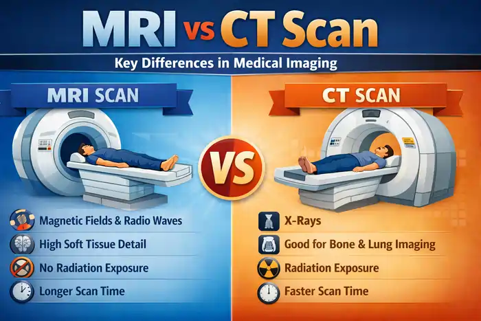
- Technology used
CT scans use X‑rays and computer processing, while MRI scans rely on magnetic fields and radio waves. - Radiation exposure
CT scans involve exposure to ionizing radiation, while MRI scans do not use radiation. Safety information from the FDA and CDC explains that medically necessary CT scans are considered safe when performed with optimized radiation doses, as discussed in their patient imaging safety resources. - Best use cases
CT scans are often used for trauma injuries, internal bleeding, lung disease, and bone fractures.
MRI scans are often used for brain disorders, spinal cord injuries, ligament injuries, and soft tissue tumors. - Image detail
MRI generally provides greater soft‑tissue detail, while CT scans are better for bone structures and rapid emergency imaging. Radiology practice guidelines often recommend CT first in acute trauma, followed by MRI for deeper soft-tissue evaluation when needed.
When Doctors Recommend a CT Scan
Healthcare providers may recommend CT imaging in several situations. Common reasons include:
- evaluating trauma injuries
- diagnosing internal bleeding
- detecting lung disease
- identifying kidney stones
- diagnosing appendicitis
- evaluating cancer spread
Because CT scans are fast and widely available, they are frequently used in emergency departments. Evidence-based emergency care protocols, such as those referenced in stroke and trauma guidelines and summarized by national health agencies, explain when CT is preferred for rapid assessment in life-threatening situations.
When Doctors Recommend an MRI
MRI scans are typically recommended when detailed imaging of soft tissues is required. Common uses include:
- brain tumors
- stroke evaluation
- spinal cord injuries
- ligament tears
- joint injuries
- multiple sclerosis diagnosis
MRI is also commonly used for detecting abnormalities in organs such as the liver, pancreas, and reproductive organs. Clinical recommendations from neurology, oncology, and orthopedic societies, which are often cited alongside NIH‑funded imaging studies, describe how MRI supports more precise diagnosis for conditions like multiple sclerosis, soft‑tissue tumors, and cartilage injuries.
Safety Considerations
Both MRI and CT scans are generally safe when used appropriately.
CT Scan Safety
CT scans involve radiation exposure, but the amount used in medical imaging is carefully controlled. Public health guidance from the CDC and regulatory information from the FDA emphasize that the diagnostic benefits of CT imaging often outweigh the potential risks when scans are medically necessary, and they encourage patients to discuss imaging history and risk with clinicians using consumer-friendly resources on radiation in medical imaging.
MRI Safety
MRI scans do not involve radiation. However, the strong magnetic field means patients with certain implants may not be eligible for MRI. Examples include:
- pacemakers
- cochlear implants
- metal fragments in the body
Healthcare providers screen patients carefully before MRI scans to ensure safety. MRI safety recommendations from radiology societies and device manufacturers, often referenced by NIH and WHO, provide detailed criteria for MRI‑safe and MRI‑conditional implants.
Preparing for MRI or CT Scans
Preparation for imaging tests varies depending on the procedure. Patients may be asked to:
- remove metal objects
- avoid food or drink for several hours
- drink contrast dye
- wear hospital gowns
Some imaging procedures use contrast agents, which help highlight blood vessels and tissues for clearer images. For guidance on preparing for laboratory and diagnostic tests—including fasting, medication timing, and what to expect before imaging—you can read our guide on how to prepare for lab testing.
Imaging Tests and Preventive Medicine
Diagnostic imaging also plays an important role in preventive healthcare. Examples include:
- CT lung cancer screening for high‑risk individuals
- cardiac CT scans to assess coronary artery disease
- MRI scans for early detection of certain neurological conditions
Preventive imaging strategies are reflected in recommendations from expert groups such as the U.S. Preventive Services Task Force and major cardiology and oncology societies, which are frequently referenced in NIH and CDC overviews of screening and early detection programs.
Imaging vs Laboratory Tests
Medical imaging and laboratory tests often work together to diagnose disease. For example:
- blood tests may detect abnormal biomarkers
- imaging scans can identify structural changes in organs
If you want to understand how blood testing works, read our article on understanding blood tests (CBC, lipid panel, A1C). Combining laboratory testing with imaging allows doctors to make more accurate diagnoses by correlating structural changes with lab findings.
Advances in Medical Imaging
Technological innovation continues to improve medical imaging. Emerging developments include:
- artificial intelligence in radiology
- high‑resolution 3D imaging
- functional MRI (fMRI) for brain activity
- molecular imaging for cancer detection
The NIH highlights advanced imaging technologies and AI‑assisted interpretation as major focuses of biomedical research, with programs exploring AI for faster scan reading, improved lesion detection, and more personalized treatment planning, as described in their biomedical imaging and engineering initiatives. International imaging societies are also publishing standards for integrating AI into radiology while maintaining quality and patient safety.
Key Takeaways | MRI vs CT Scan
MRI and CT scans are essential diagnostic imaging tools that help doctors detect disease, evaluate injuries, and monitor treatment outcomes. CT scans use X‑rays to create cross-sectional images and are commonly used in emergency medicine and trauma evaluation. MRI scans use magnetic fields and radio waves to produce highly detailed images of soft tissues, making them especially useful for neurological and musculoskeletal conditions. Understanding the differences between these imaging tests can help patients feel more informed and prepared when undergoing diagnostic procedures.
Medical Disclaimer
This article is intended for educational purposes only and should not replace professional medical advice, diagnosis, or treatment. Always consult a qualified healthcare professional for personalized medical guidance regarding imaging tests, health conditions, or treatment decisions.
Written by: Eden Grace Ramos, RN
Medical Resources
This article references evidence-based guidance from trusted health authorities to ensure clinical accuracy and reliability. Key resources include the National Institutes of Health medical imaging research programs, public health guidance from the Centers for Disease Control and Prevention and U.S. Food and Drug Administration on medical imaging and radiation safety, and global healthcare guidance from the World Health Organization on diagnostic imaging and health systems. These organizations provide research and clinical recommendations widely used by healthcare professionals worldwide.