Basic Neuroanatomy Principles Key Takeaways
Whether you are a medical student wrestling with your first brain dissection or a resident preparing for board exams, the basic neuroanatomy principles are your map.
- Basic Neuroanatomy Principles follow a logical hierarchy: the nervous system is organized from central to peripheral, with distinct functional systems that cross anatomical boundaries.
- Understanding the brain structure organization and spinal cord anatomy basics makes it easier to grasp complex clinical syndromes like stroke localization and spinal cord injury patterns.
- Effective neuroanatomy memorization techniques rely on spatial relationships, functional pathways, and repeated practice with real clinical cases rather than rote list learning.
Why the Basic Neuroanatomy Principles Matter for Clinical Practice
Whether you are a medical student wrestling with your first brain dissection or a resident preparing for board exams, the basic neuroanatomy principles are your map. These nine rules will help you navigate the complex terrain of the central nervous system anatomy without getting lost. By internalizing these neuroanatomy basics, you lay the groundwork for understanding brain structure organization, spinal cord anatomy basics, and the nervous system structure as a whole. For a related guide, see 10 Essential Neurology Concepts Every Medical Student Must Know.

Why is neuroanatomy important for medical students? Because every neurological symptom—from a drooping eyelid to a gait disturbance—originates in a specific anatomical structure. The basic neuroanatomy principles give you the language to describe what is happening and where. When you learn neuroanatomy through these principles, you move beyond memorization into genuine clinical reasoning.
Principle 1: The Nervous System Has Two Major Anatomical Divisions
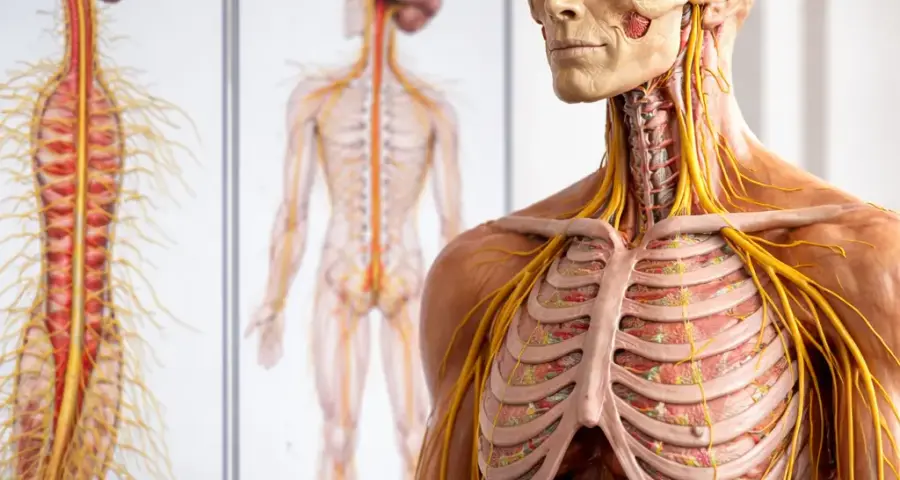
Before diving into any brain region, commit this fundamental rule to memory: the nervous system organization principles begin with the division between the central nervous system (CNS) and the peripheral nervous system (PNS). The CNS consists of the brain and spinal cord, while the PNS comprises all nerves outside the CNS. This is the most basic of the basic neuroanatomy principles. For a related guide, see Neurological Conditions: Symptoms, Signs, and Treatment.
Understanding CNS and PNS Differences
The CNS processes and integrates information; the PNS carries sensory input to the CNS and motor commands away from it. A common mistake in learning neuroanatomy is treating these as separate systems, but they function as a continuous loop. For example, when you touch a hot surface, sensory nerves (PNS) send the signal to your spinal cord (CNS), which then sends a motor command back through the PNS to pull your hand away.
Clinical relevance: Understanding CNS and PNS differences helps you localize lesions. A patient with weakness but normal reflexes may have a peripheral nerve problem, while one with spasticity and hyperreflexia likely has a CNS lesion.
Memorization tip: Use the mnemonic “CNS is Command Central; PNS is the Postal Service.”
Principle 2: The Brain and Spinal Cord Are Organized Along a Rostral-Caudal Axis
The brain and spinal cord structure runs from the top (rostral) to the bottom (caudal). This axis is critical for understanding how brain regions functions are arranged. The brain and spinal cord anatomy follows a predictable pattern: from the cerebrum down through the brainstem to the spinal cord.
Neural Pathways Organization Along the Axis
Major neural pathways organization—such as the corticospinal tract and the spinothalamic tract—travel along this axis. Understanding the rostral-caudal arrangement helps you predict what deficits will occur with lesions at different levels. This is why brain mapping basics always start with orientation along this axis.
Clinical relevance: A stroke in the left cerebral hemisphere (rostral) may cause right-sided weakness, while a spinal cord injury in the cervical region (caudal) can cause weakness in all four limbs.
Memorization tip: Picture the CNS as a vertical column. Draw it from top to bottom in your notes every day for a week.
Principle 3: The Brain Has Three Major Structural and Functional Divisions
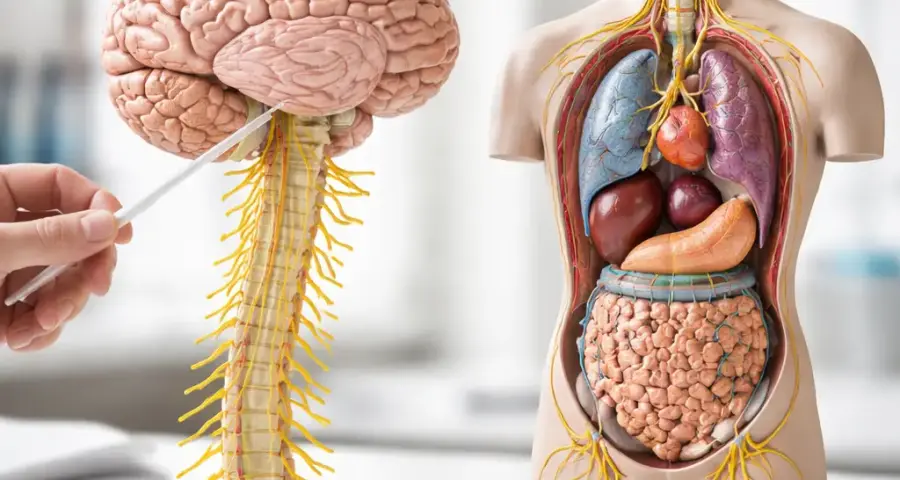
These are the cerebrum, the brainstem, and the cerebellum. This brain structure organization principle helps you group functions: the cerebrum handles higher cognition, the brainstem controls basic life functions, and the cerebellum coordinates movement. This is one of the most important functional neuroanatomy concepts.
Brain Lobes and Functions
Each cerebral hemisphere is divided into four lobes: frontal, parietal, temporal, and occipital. The brain lobes and functions map follows a general pattern: frontal for motor control and executive function, parietal for sensation, temporal for memory and hearing, and occipital for vision. Understanding this anatomical brain relationships framework is essential for clinical neuroanatomy introduction.
Clinical relevance: A tumor in the frontal lobe may cause personality changes; one in the occipital lobe may cause vision loss.
Memorization tip: Create a color-coded diagram of the lobes. Draw it without labels first, then add them one by one.
Principle 4: Gray Matter Contains Neuron Cell Bodies; White Matter Contains Axons
Understanding white matter and gray matter basics is non-negotiable. Gray matter appears gray in fresh specimens because it contains neuron cell bodies, dendrites, and unmyelinated axons. White matter appears white because of the myelin sheaths that insulate axons and speed up signal transmission. This is a cornerstone of medical neuroanatomy fundamentals.
Where Gray and White Matter Live
In the brain, gray matter forms the outer cortex and internal nuclei like the thalamus and basal ganglia. White matter makes up the tracts that connect different brain regions. In the spinal cord anatomy basics, the pattern reverses: gray matter forms a butterfly-shaped interior, while white matter surrounds it. This reversal is a common point of confusion for beginners.
Clinical relevance: Multiple sclerosis attacks white matter, causing demyelination and an array of symptoms depending on which tracts are affected.
Memorization tip: Think “Gray matter is the town; white matter is the roads connecting towns.”
Principle 5: The Spinal Cord Has a Consistent Segmental Organization
The spinal cord anatomy basics follow a segmental pattern: cervical, thoracic, lumbar, and sacral. Each segment gives rise to a pair of spinal nerves that carry both sensory and motor fibers. This nervous system structure principle allows you to map symptoms to a specific spinal level.
Neural Pathways Organization in the Cord
Key pathways like the corticospinal tract (motor) and the spinothalamic tract (pain and temperature) run through specific regions of the white matter. Knowing their location helps you predict nervous system structure function relationships. For example, the corticospinal tract is in the lateral funiculus; damage there causes ipsilateral weakness below the lesion.
Clinical relevance: A C5 spinal cord injury affects the arms and legs; an L1 injury affects only the legs.
Memorization tip: Use dermatome maps. Learn the key landmarks: C6 (thumb), T4 (nipple line), T10 (umbilicus), L4 (knee).
Principle 6: The Cranial Nerves Are the Special Messengers of the Head and Neck
There are 12 pairs of cranial nerves. This cranial nerves overview is essential because each nerve has a specific function—sensory, motor, or both—and emerges from specific brainstem nuclei. This is a vital component of clinical neuroanatomy introduction for anyone learning neuroanatomy for beginners.
How to Learn Cranial Nerves Efficiently
Group them by function: CN I and II are purely sensory (smell and vision); CN III, IV, VI, XI, and XII are mostly motor; CN V, VII, IX, and X are mixed. Many neuroanatomy memorization techniques rely on mnemonics like “Oh, Oh, Oh, To Touch And Feel Very Good Velvet, Ah, Heaven” to remember the names.
Clinical relevance: A patient with a head turn weakness has a CN XI (spinal accessory) problem; facial drop with sparing of the forehead suggests a central (upper motor neuron) lesion of CN VII.
Memorization tip: Test yourself by drawing a skull and placing each nerve at the correct exit point. Do it from memory, then check.
Principle 7: The Ventricular System and Meninges Protect the CNS
The brain and spinal cord structure is enclosed by three meningeal layers: dura mater, arachnoid mater, and pia mater. Inside the brain, a system of interconnected cavities—the ventricles—produces and circulates cerebrospinal fluid (CSF). This medical neuroanatomy fundamentals principle is often tested in exams.
Clinical Anatomy of Brain Protection
The clinical anatomy of brain protection matters because bleeding between these layers has distinct clinical patterns. An epidural hematoma lies above the dura, a subdural hematoma lies below it, and a subarachnoid hemorrhage occurs between the arachnoid and pia. Each has a characteristic presentation and imaging appearance.
Clinical relevance: A patient on anticoagulants with a headache and altered consciousness may have a subdural hematoma.
Memorization tip: Remember the layers from outside to inside: “Dura, Arachnoid, Pia” → “DAP.” For ventricles: “Lateral, Third, Aqueduct, Fourth” → “LTAF.”
Principle 8: The Blood Supply to the Brain Comes from Two Major Systems
The internal carotid arteries (anterior circulation) and the vertebral arteries (posterior circulation) supply the brain. They anastomose at the Circle of Willis at the base of the brain. Understanding this brain structure organization principle is critical for brain mapping basics and stroke localization.
Brain Structure Function Relationship in Vascular Territories
The anterior circulation supplies most of the cerebrum; the posterior circulation supplies the brainstem, cerebellum, and occipital lobes. Knowing which artery supplies which region helps you predict deficits after an occlusion. This is one of the most practical basic neuroanatomy principles for clinicians.
Clinical relevance: Middle cerebral artery stroke causes contralateral weakness and sensory loss, plus aphasia if the dominant hemisphere is affected.
Memorization tip: Draw the Circle of Willis as a hexagon. Label each vessel and the region it supplies. Then test yourself with “bleeding” scenarios.
Principle 9: The Nervous System Uses Both Ipsilateral and Contralateral Pathways
Many major pathways cross (decussate) at some point along the neuraxis. For example, the corticospinal tract crosses at the medullary pyramids, so the left brain controls the right side of the body. The dorsal column-medial lemniscus pathway (fine touch and proprioception) crosses in the medulla as well. The spinothalamic tract (pain and temperature) crosses in the spinal cord near its entry level. This neural pathways organization principle explains why lesions produce specific patterns of deficits.
How This Affects Neuroanatomy Basics for Clinical Reasoning
When you understand crossing patterns, you can localize a lesion based on symptom distribution. For example, a patient with left-sided pain and temperature loss but right-sided weakness likely has a lesion on the right side of the spinal cord or lower brainstem. This is a classic application of neurophysiology basics combined with anatomy.
Clinical relevance: Brown-Séquard syndrome (incomplete spinal cord injury) presents with ipsilateral weakness and loss of proprioception, plus contralateral pain and temperature loss below the lesion.
Memorization tip: Memorize the three main crossing rules: motor crosses in the medulla; fine touch crosses in the medulla; pain/temperature crosses at the spinal entry level.
How to Learn Neuroanatomy Easily: A Practical Framework
How do I learn neuroanatomy easily? Start with these basic neuroanatomy principles and build layer by layer. Many learners make the mistake of trying to memorize every structure at once. Instead, use the following approach:
- Master the orientation: Learn the planes (coronal, sagittal, horizontal) and directions (rostral, caudal, ventral, dorsal).
- Learn the major divisions: CNS vs. PNS, brain vs. brainstem vs. cerebellum.
- Add the ventricles and meninges: The “container” comes before the “contents.”
- Study major pathways one at a time: Corticospinal tract, then spinothalamic tract, then dorsal columns.
- Apply to clinical vignettes: For each pathway, ask: “Where would a lesion cause this patient’s symptoms?”
Common mistakes in learning neuroanatomy include skipping the spatial relationships, trying to learn from text alone, and memorizing names without understanding connections. Use neuroanatomy study tips like drawing, labeling, and teaching another student to solidify your knowledge.
What are the key structures in neuroanatomy? Focus on these core structures: cerebrum with its lobes, brainstem (midbrain, pons, medulla), cerebellum, spinal cord segments, cranial nerves, basal ganglia, thalamus, hypothalamus, and the major white matter tracts. These are the pillars of medical neuroscience foundations.
Useful Resources
To reinforce your understanding of these basic neuroanatomy principles, explore these authoritative resources:
- University of Wisconsin Neuroanatomy Laboratory — A well-regarded resource with interactive atlases, dissection guides, and self-assessment quizzes designed for medical and neuroscience students.
- Radiopaedia Neuroanatomy Articles — A trusted clinical radiology site offering detailed anatomical descriptions, imaging correlations, and pathology examples that bring the basic neuroanatomy principles to life in a practical context.
Mastering the basic neuroanatomy principles is not an overnight task, but with consistent practice, drawing, and clinical application, you will develop the spatial intuition needed to excel in medical neuroscience and patient care. Keep returning to these nine principles whenever you feel lost—they are your compass through the complexity of the human nervous system.
Frequently Asked Questions About Basic Neuroanatomy Principles
What are the basic principles of neuroanatomy?
The basic principles include division of the nervous system into CNS and PNS, rostral-caudal organization, gray vs. white matter differences, segmental organization of the spinal cord, cranial nerve functions, the ventricular and meningeal protection system, dual blood supply, and the crossing of major pathways. These basic neuroanatomy principles form the foundation for clinical localization.
Why is neuroanatomy important for medical students?
Neuroanatomy is the road map for diagnosing neurological conditions. Every symptom—weakness, numbness, vision loss, dizziness—points to a specific anatomical structure. Without understanding neuroanatomy basics, medical students cannot accurately localize lesions or interpret neurological exam findings. Mastery of basic neuroanatomy principles directly impacts patient care and board exam performance.
How do I learn neuroanatomy easily?
Learn neuroanatomy by focusing on brain structure organization first: orientation planes, major divisions, and key landmarks. Use active learning: draw diagrams, label structures, teach a peer, and apply knowledge to clinical cases. Spaced repetition with flashcards for cranial nerves overview and spinal cord anatomy basics helps retention. Avoid passive reading.
What are the key structures in neuroanatomy?
The key structures include the cerebrum (with frontal, parietal, temporal, and occipital lobes), brainstem (midbrain, pons, medulla), cerebellum, spinal cord, thalamus, hypothalamus, basal ganglia, limbic system, cranial nerves 1–12, and major white matter tracts. Understanding brain regions functions and their connections is central to medical neuroanatomy fundamentals.
What are the fundamental rules of brain organization?
The fundamental rules include the rostral-caudal organization of the CNS, separation of gray and white matter, division into functional lobes with specific functions, the crossing of most sensory and motor pathways, and the dual arterial supply from carotid and vertebral systems. These nervous system organization principles govern how all brain regions interact.
How does the nervous system structure function?
The nervous system uses the CNS (brain and spinal cord) to process information and the PNS to collect sensory input and execute motor output. The nervous system structure function relationship is hierarchical: the cerebrum plans, the brainstem modulates vital functions, and the spinal cord executes reflexes. Communication occurs via neural pathways organization of ascending and descending tracts. For a related guide, see 12 Key Steps in the Neurological Examination Explained Simply.
What are the basics of brain and spinal cord anatomy?
The brain consists of cerebrum, brainstem, and cerebellum; the spinal cord is a segmented tube of gray matter surrounded by white matter. The brain and spinal cord structure is protected by meninges and CSF. Key spinal cord anatomy basics include the butterfly-shaped gray matter and the three funiculi of white matter containing major pathways.
How can I memorize neuroanatomy effectively?
Use neuroanatomy memorization techniques such as drawing and labeling structures from memory, employing mnemonics (for cranial nerves, for example), creating story-based associations, and practicing with clinical vignettes. Spaced repetition software for brain anatomy study tips is also highly effective. The goal is to build spatial and functional understanding, not just a list of names.
What are common mistakes in learning neuroanatomy?
Common mistakes include trying to memorize every small structure without understanding brain structure organization, ignoring the rostral-caudal axis, confusing white and gray matter locations, failing to learn pathways including their crossing points, and skipping the meningeal and ventricular systems. These mistakes lead to confusion in clinical neuroanatomy introduction and exam performance.
Why is neuroanatomy difficult for beginners?
Neuroanatomy is challenging because of the density of structures, the complex three-dimensional organization of central nervous system anatomy, and the need to integrate spatial understanding with function. Beginners often feel overwhelmed by the volume of terminology. Starting with basic neuroanatomy principles and building gradually reduces this difficulty.
What is the best way to study brain lobes and functions ?
Study brain lobes and functions by linking each lobe to a major clinical syndrome: frontal lobe to executive dysfunction, parietal to neglect, temporal to memory loss, occipital to visual deficits. Draw a lateral view of the brain, label the lobes, and write one key function and one clinical sign for each. Repeat until you can do it from memory.
How do cranial nerves connect to brainstem nuclei?
Each cranial nerve originates from or terminates in specific brainstem nuclei. For example, the oculomotor nerve (CN III) originates from the oculomotor nucleus in the midbrain. The cranial nerves overview should include the name, number, function, nucleus location, and exit point from the skull for each nerve.
What is the role of white matter in the brain?
White matter contains the axons of neurons, which are bundled into tracts that connect different gray matter regions. White matter and gray matter basics teach that white matter is essential for communication between brain areas. Damage to white matter disrupts these connections, leading to disconnection syndromes.
How is the spinal cord organized segmentally?
The spinal cord is divided into cervical, thoracic, lumbar, and sacral segments. Each segment gives off a pair of spinal nerves. Spinal cord anatomy basics emphasize that the number of segments (31: 8 cervical, 12 thoracic, 5 lumbar, 5 sacral, 1 coccygeal) corresponds to the vertebrae but with offset in adults.
What are the major neural pathways in the CNS?
Major pathways include the corticospinal tract (motor), dorsal column-medial lemniscus (fine touch/vibration), spinothalamic tract (pain/temperature), and spinocerebellar tracts (proprioception to cerebellum). Neural pathways organization describes the path from origin to termination, including crossing points.
Why is the Circle of Willis important?
The Circle of Willis is an anastomotic ring at the base of the brain that connects the carotid and vertebral circulations. It provides collateral blood flow, so if one vessel is blocked, blood can reach brain regions via alternate routes. This brain structure organization principle is key in stroke risk and management.
What is the clinical significance of the corticospinal tract crossing?
Because the corticospinal tract crosses in the medulla, a lesion above the crossing (in the brain or above the medulla) produces contralateral weakness. A lesion below the crossing (in the spinal cord) produces ipsilateral weakness. This neural pathways organization fact is essential for lesion localization.
How do I differentiate between upper and lower motor neuron lesions?
Upper motor neuron lesions cause spasticity, hyperreflexia, and Babinski sign. Lower motor neuron lesions cause flaccidity, hyporeflexia, and muscle atrophy. Understanding the pathway from cortex to muscle helps you apply basic neuroanatomy principles to differentiate them.
What are the ventricles and what do they do?
The brain contains four ventricles: two lateral ventricles, the third ventricle, and the fourth ventricle. They produce and circulate cerebrospinal fluid, which cushions the CNS, removes waste, and maintains pressure. This medical neuroanatomy fundamentals topic is often tested in relation to hydrocephalus.
What is the best resource for neuroanatomy review?
Excellent resources include Nolte’s “The Human Brain,” Snell’s “Clinical Neuroanatomy,” the Neuroanatomy section of Radiopaedia, and the University of Wisconsin online neuroanatomy lab. Combining textbook reading with brain mapping basics from these resources reinforces neuroanatomy basics effectively.