
Simple Ways to Localize Neurological Lesions Key Takeaways
Neurological localization—the process of identifying where in the central or peripheral nervous system a lesion is located—is the cornerstone of every accurate neurologic diagnosis.
- Simple Ways to Localize Neurological Lesions means starting with the history and exam, then applying pattern recognition to pinpoint the site of damage.
- Understanding upper motor neuron vs lower motor neuron lesions forms the first key branching point in any localization algorithm.
- Combining motor sensory pathway localization with cranial nerve findings often narrows the lesion to a specific CNS region.
What Does Neurological Lesion Localization Mean?
Neurological lesion localization is the clinical art and science of determining the precise anatomic site of a pathologic process within the nervous system. It is the first and most critical step in every neurologic consultation. Instead of simply listing symptoms, the clinician asks: Where is the lesion causing these symptoms?

This process relies on functional neuroanatomy localization—knowledge of what each structure does and what deficits arise when it is damaged. For example, a patient who presents with right-sided weakness and speech difficulty likely has a left hemisphere lesion. A patient with crossed signs (ipsilateral face, contralateral body) points toward a brainstem lesion. These patterns are the building blocks of clinical neuro localization. For a related guide, see 10 Red Flags in Neurological Assessment You Should Never Miss.
How Do I Localize Neurological Lesions Step by Step?
Lesion localization step by step follows a logical sequence that narrows the possible sites from general to specific. Here is a structured approach used in clinical neurology training:
Step 1: Define the Problem
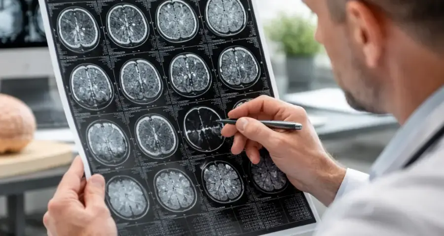
Start with the history. Is the weakness acute (stroke) or chronic (tumor)? Is the numbness ascending (Guillain-Barré) or dermatomal (nerve root)? This immediately suggests a category of pathology and narrows the anatomic possibilities.
Step 2: Distinguish UMN vs LMN
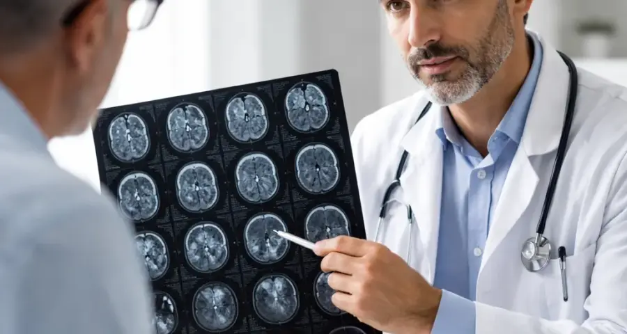
Determine whether the lesion involves the upper motor neuron (brain or spinal cord) or lower motor neuron (anterior horn cell, nerve root, peripheral nerve). Upper motor neuron vs lower motor neuron lesions have distinct features: UMN causes spasticity, hyperreflexia, and a Babinski sign; LMN causes flaccidity, hyporeflexia, and atrophy. This single step eliminates half the nervous system. For a related guide, see Neurological Conditions: Symptoms, Signs, and Treatment.
Step 3: Map the Sensory and Motor Levels
If the lesion is supratentorial (above the brainstem), expect hemiparesis or hemisensory loss on the contralateral side. If the lesion is below the neck, a spinal cord level can be established by finding a sensory transition zone on the trunk and assessing for bowel/bladder involvement.
Step 4: Check the Cranial Nerves
Cranial nerve findings are powerful localizers. A third nerve palsy with contralateral hemiparesis (Weber syndrome) localizes to the midbrain. A lateral medullary syndrome (Wallenberg) presents with ipsilateral Horner, ataxia, and facial numbness with contralateral body pain/temperature loss. Recognizing these brainstem lesion signs is essential for brainstem lesion diagnosis.
What Are the Key Rules for Lesion Site Determination?
Lesion site determination is guided by several core neuroanatomy localization principles that every clinician should memorize:
- Rule of decussation: Motor pathways cross at the medullary pyramids; sensory pathways cross at the spinal cord level. Lesions above the decussation produce contralateral deficits; lesions below produce ipsilateral deficits (except in the face for certain pathways).
- Rule of the spinal cord level: A transverse myelopathy causes bilateral motor and sensory loss at and below a specific spinal segment, often with sphincter involvement. A Brown-Séquard syndrome (hemisection) causes ipsilateral motor loss and contralateral pain/temperature loss.
- Rule of the brainstem: Cranial nerve nuclei are located here. A lesion affecting a cranial nerve on one side and the motor/sensory tracts on the opposite side always indicates a brainstem localization (the so-called “crossed” or “alternating” syndrome).
- Rule of cortical representation: The primary motor and sensory cortices are somatotopically organized. A lesion in the leg area of the motor homunculus causes leg-predominant weakness; a lesion in the face area causes face-predominant weakness. This is the basis for cortical lesion identification.
How Can I Identify Lesion Sites in the Brain or Spinal Cord?
Distinguishing brain vs spinal cord lesions is a frequent clinical challenge. Here are practical CNS lesion differentiation criteria:
| Feature | Brain Lesion | Spinal Cord Lesion |
|---|---|---|
| Face involvement | Common (especially with hemispheric or brainstem lesions) | Absent (unless lesion is very high cervical, but then also affects neck) |
| Hemiparesis distribution | Face, arm, leg on one side | Bilateral below a level; arms can be spared in thoracic lesions |
| Sensory signs | Hemisensory loss (same side as motor) | Dermatomal level; sometimes dissociated (pain/temp vs touch/vibration) |
| Bowel/bladder | Often spared early; can be affected with bilateral lesions | Early and common |
| Cranial nerve signs | Present with brainstem lesions; absent with pure cord lesions | Absent (below the foramen magnum) |
This table helps you perform brain and spinal cord lesion diagnosis quickly at the bedside.
What Clinical Signs Help in Neurological Exam Interpretation?
Mastering neurological exam interpretation means knowing which signs point directly to a specific structure. Here are the most clinically useful patterns:
Motor Pathway Lesion Signs
When you find spasticity, hyperreflexia, and a Babinski sign, you have localized the lesion to the upper motor neuron. The question then becomes: is it in the hemisphere, brainstem, or spinal cord? Additional signs like aphasia, hemineglect, or visual field cut point to a cortical or subcortical hemisphere lesion. If the motor findings are bilateral below a spinal level, the lesion is in the cord.
Sensory Pathway Lesion Patterns
Sensory pathway lesion patterns can be even more precise. A lesion in the dorsal columns (posterior cord) causes loss of vibration and proprioception below the lesion. A lesion in the spinothalamic tract causes contralateral loss of pain and temperature. Recognizing these patterns allows you to differentiate between a cord lesion (level) and a brainstem lesion (crossed findings).
Cranial Nerve Lesion Patterns
Cranial nerve lesion patterns are among the most reliable localizers. For example:
- CN III palsy + contralateral hemiparesis = midbrain (Weber syndrome)
- CN VI palsy + ipsilateral facial weakness + contralateral hemiparesis = pons (Millard-Gubler syndrome)
- CN VIII (hearing loss, vertigo) + ipsilateral facial numbness + ataxia = cerebellopontine angle (acoustic neuroma)
These syndromes are classic examples of cranial nerve examination localization in action.
Why Is Lesion Localization Important in Diagnosis?
Lesion localization is important because it directly determines the differential diagnosis. A stroke in the basal ganglia has a different treatment and prognosis than a demyelinating plaque in the spinal cord. An anterior horn cell lesion (ALS) demands a different workup than a peripheral neuropathy. Without localization, imaging and lab tests become shots in the dark. With precise localization, you can order the right MRI sequence, the right lab test, or the right electrophysiology study.
Moreover, localization often happens before any imaging. A skilled clinician can say, “The lesion is in the left midbrain,” and the MRI confirms it. This saves time, reduces unnecessary testing, and builds confidence in your neuro exam pattern recognition.
What Are Common Patterns in Neuro Lesion Localization?
Neuro lesion localization follows recognizable patterns that form the basis of neuroanatomy case based learning. Here are some classic scenarios:
Hemispheric Syndrome
Unilateral weakness and sensory loss, often with higher cortical dysfunction (aphasia, neglect, apraxia). The lesion is in the contralateral hemisphere. Specific homunculus patterns can localize to the motor or sensory strip.
Brainstem Syndrome
Crossed signs (ipsilateral cranial nerve, contralateral body motor/sensory). This is the hallmark of a brainstem lesion. Combinations of cranial nerve palsies and long tract signs point to a specific level (midbrain, pons, medulla).
Spinal Cord Syndrome
Bilateral motor and sensory loss below a defined level, with sphincter involvement. Variants include:
- Complete transection: bilateral paralysis, sensory loss, autonomic loss
- Brown-Séquard: ipsilateral motor, contralateral pain/temp loss
- Central cord: more weakness in arms than legs, sacral sparing
- Anterior cord: paralysis and pain/temp loss, preserved proprioception
Peripheral Nerve Syndrome
Mononeuropathy follows a specific nerve distribution (e.g., median, ulnar, radial). Polyneuropathy is length-dependent, starting in the feet and ascending.
How Do Motor and Sensory Findings Help Localize Lesions?
Motor sensory pathway localization is one of the most powerful tools in clinical neuroanatomy reasoning. Here is how to combine the two:
- If a patient has weakness and sensory loss affecting the same side of the body (face, arm, leg), the lesion is in the contralateral hemisphere or brainstem.
- If the weakness and sensory loss affect both legs (paraplegia) with a sensory level on the trunk, the lesion is in the thoracic or lumbar spinal cord.
- If the weakness and sensory loss are in one arm only, the lesion could be in the contralateral motor cortex (focal) or in the cervical roots/plexus/peripheral nerve.
- If the weakness is unilateral but sensory loss is minimal, suspect a pure motor stroke (lacunar infarct) in the internal capsule or pons.
- If the sensory loss is dissociated (pain/temp lost, vibration/proprioception spared), the lesion is in the spinothalamic tract, usually in the spinal cord or brainstem.
These motor pathway lesion signs and sensory pathway lesion patterns form the core of your localization toolkit.
8 Simple Ways to Localize Neurological Lesions
This is the practical neurological localization guide you have been waiting for. Each method builds on the previous one to help you perform brain lesion mapping and spinal cord injury localization with confidence.
Method 1: Start with the History
Always begin by asking: What did the patient feel first? A sudden onset suggests vascular or traumatic injury. A slow, progressive course suggests tumor, infection, or degenerative disease. An intermittent pattern could be multiple sclerosis or transient ischemic attack. The history also tells you about the speed of symptom evolution, which helps narrow the pathology type.
Method 2: Perform a Targeted Neurologic Exam
Focus on motor strength, reflexes, tone, sensation (light touch, pinprick, vibration, proprioception), coordination, and cranial nerves. Use a systematic approach that covers all major CNS lesion identification domains. Always examine gait if the patient can walk.
Method 3: Differentiate UMN vs LMN
This is the single most powerful dichotomy in localization. UMN signs (spasticity, hyperreflexia, Babinski) point to the brain or spinal cord. LMN signs (flaccidity, hyporeflexia, atrophy, fasciculations) point to the anterior horn cell, nerve root, or peripheral nerve. This immediately halves the number of possible lesion sites.
Method 4: Determine the Laterality
Is the deficit unilateral, bilateral, or symmetric? Unilateral hemisphere or brainstem lesions cause contralateral findings. Bilateral hemisphere lesions (e.g., Bickerstaff encephalitis) are rare. Bilateral spinal cord lesions cause paraplegia or quadriplegia with a sensory level. This step helps you differentiate brain vs spinal cord lesions.
Method 5: Look for a Sensory Level
Use a cotton wisp or pin to map the sensory loss from the neck down. A distinct transition (e.g., below the T4 dermatome) indicates a spinal cord lesion at that segment. A complete lack of a level suggests a brain lesion or peripheral neuropathy.
Method 6: Analyze Cranial Nerve Findings
As noted earlier, cranial nerve lesion patterns are key localizers. Pay special attention to CN II (visual loss, field defects), CN III (pupil, extraocular movements), CN VII (facial weakness—upper vs lower motor neuron), and CN XII (tongue deviation). A lower motor neuron facial palsy (Bell palsy) spares the forehead; an upper motor neuron lesion (stroke) typically affects the lower face only.
Method 7: Use Reflex Changes
Hyperreflexia localizes to the upper motor neuron. Hyporeflexia or areflexia localizes to the lower motor neuron, nerve root, or peripheral nerve. Pathologic reflexes (Babinski, Hoffman, clonus) are always upper motor neuron signs. Also test the jaw jerk—an exaggerated jaw jerk indicates a lesion above the pons (pseudobulbar palsy).
Method 8: Integrate with Imaging and Labs
Finally, confirm your clinical localization with appropriate neuro diagnostic reasoning tools: MRI with contrast for brain or spinal cord, CT for acute stroke, EMG/NCS for peripheral nerve or muscle, and lumbar puncture if infection or inflammation is suspected. Your clinical localization tells you where to look; imaging confirms it.
Clinical Neurology Localization Techniques in Action
Here is a case example to show these clinical neurology localization techniques at work:
Case: A 68-year-old man presents with acute onset left arm and leg weakness. On exam, he has left-sided spastic hemiparesis, hyperreflexia, and a left Babinski sign. He also has a left visual field cut and mild dysarthria.
Analysis:
- UMN signs (spasticity, Babinski) → lesion in brain or spinal cord, not LMN.
- Unilateral deficit → contralateral hemisphere or brainstem, not cord (no bilateral signs).
- Visual field cut → lesion involves the optic radiations, which pass through the temporal and parietal lobes. This localizes to the right hemisphere.
- Dysarthria without aphasia → helps rule out dominant hemisphere.
- Final localization: Right hemisphere lesion, likely involving the motor cortex and optic radiations, consistent with an ischemic stroke in the middle cerebral artery territory.
This is a classic example of applying brain region damage signs to arrive at a precise diagnosis.
Neurological Diagnosis Techniques for Common Scenarios
Here are quick reference neurological diagnosis techniques for common presentations:
| Presentation | Likely Location | Key Confirming Signs |
|---|---|---|
| Sudden onset unilateral weakness + aphasia | Left hemisphere (dominant) | Right hemiparesis, non-fluent (Broca) or fluent (Wernicke) aphasia |
| Crossed signs: right face weakness + left body weakness | Left brainstem (pons/medulla) | Cranial nerve VII or XII on right, motor tract on left |
| Paraplegia + sensory level at T10 + bladder retention | Thoracic spinal cord (T10 lesion) | Bilateral UMN signs, sphincter involvement, dermatomal sensory level |
| Diplopia, ptosis, dilated pupil on one side | CN III nucleus or nerve (midbrain or cavernous sinus) | Unilateral CN III palsy, possible midbrain signs |
| Progressive leg weakness and numbness, absent knee and ankle jerks | Peripheral neuropathy (LMN) | Distal weakness, stocking-glove sensory loss, areflexia |
These lesion site determination shortcuts are invaluable at the bedside.
Useful Resources for Neurological Localization
For further study of simple ways to localize neurological lesions, we recommend these authoritative resources:
- Neuroanatomy through Clinical Cases – A widely used textbook that pairs anatomical knowledge with real clinical scenarios, ideal for neuroanatomy case based learning.
- UpToDate: Approach to the Adult with Acute Neurologic Deficit – A practical clinical algorithm for localizing acute CNS lesions, regularly updated for evidence-based care.
Frequently Asked Questions About Simple Ways to Localize Neurological Lesions
What does lesion localization in neurology mean?
Lesion localization in neurology means determining the exact anatomic site of damage within the nervous system based on clinical signs and symptoms. It is the first step in diagnosing and treating neurologic conditions.
How do I localize neurological lesions step by step?
Start with the history to define the problem, then examine for UMN vs LMN signs, map motor and sensory findings, check cranial nerves, and look for a spinal level. Use pattern recognition to narrow the lesion to a specific CNS region. This lesion localization step by step approach is systematic and reliable.
What are the key rules for lesion localization?
The key rules include: contralateral signs for brain lesions (above decussations), the crossed sign rule for brainstem lesions, a sensory level for spinal cord lesions, and the homunculus for cortical lesions. These neuroanatomy localization principles form the foundation of bedside localization.
How can I identify lesion sites in the brain or spinal cord?
Identify lesion sites by evaluating hemiparesis distribution (face-arm-leg vs paraplegia), presence of cranial nerve signs, sensory level, bowel/bladder involvement, and specific battery patterns (Brown-Séquard, central cord). This brain lesion mapping approach is highly effective.
What clinical signs help in neurological localization?
Key signs include spasticity and Babinski (UMN), flaccidity and areflexia (LMN), crossed signs (brainstem), sensory level (spinal cord), homonymous hemianopia (optic radiations), and aphasia (dominant hemisphere). These are examples of clinical neuro localization in practice.
Why is lesion localization important in diagnosis?
Lesion localization is important because it directs the differential diagnosis, helps choose the right imaging and lab tests, guides treatment decisions, and often predicts prognosis. Without it, workup becomes inefficient and potentially inaccurate.
How do motor and sensory findings help localize lesions?
Motor and sensory pathway localization helps by defining the laterality, level, and specific tract involved. Unilateral motor and sensory loss on the same side points to a contralateral brain lesion. Bilateral findings with a sensory level point to a spinal cord lesion. Dissociated sensory loss localizes to specific tracts (spinothalamic or dorsal columns).
What are common patterns in neuro lesion localization?
Common patterns include: hemisphere syndrome (contralateral hemiparesis + higher cortical signs), brainstem syndrome (crossed signs), spinal cord syndrome (bilateral deficits below a level), and peripheral nerve syndrome (single nerve distribution). Recognizing these neuro lesion localization patterns is essential for rapid diagnosis.
How do I differentiate brain vs spinal cord lesions?
Differentiate brain vs spinal cord lesions by checking for face involvement (brain), hemiparesis distribution (brain typically affects one side, cord affects both below a level), sensory level (cord), and bowel/bladder symptoms (cord). Cranial nerve signs point to brain, especially brainstem; their absence suggests cord.
What are simple methods to learn neurological localization?
The simplest methods include: learn the UMN vs LMN dichotomy first, memorize the major brainstem syndromes, practice with case studies, use a sensory mapping tool, and always ask “where is the lesion?” before “what is the lesion?”. This clinical neurology training guide approach is very effective.
What is the difference between upper motor neuron and lower motor neuron lesions?
Upper motor neuron vs lower motor neuron lesions differ in tone (spastic vs flaccid), reflexes (hyperreflexia vs hyporeflexia), and the presence of Babinski (UMN) vs fasciculations (LMN). This is the most important branching point in CNS lesion identification.
How do I recognize a brainstem lesion?
Recognize a brainstem lesion by the presence of brainstem lesion signs: ipsilateral cranial nerve palsy combined with contralateral motor or sensory loss (crossed signs). Combinations of specific cranial nerve and long tract findings point to a precise level (midbrain, pons, medulla).
How do I perform a cranial nerve examination for localization?
Cranial nerve examination localization involves testing each cranial nerve systematically. Key localizers include: CN II (visual field defects), CN III (pupil and eye movement), CN VII (upper vs lower face weakness), and CN XII (tongue deviation). Pattern recognition of cranial nerve lesion patterns is crucial.
What is a sensory level and how do I find it?
A sensory level is a horizontal line on the trunk below which sensation is diminished or absent. Find it by testing pinprick or light touch from the neck downward, noting the dermatomal level where the patient first reports a change. This is a core part of spinal cord injury localization.
What is Brown-Séquard syndrome?
Brown-Séquard syndrome is a spinal cord lesion diagnosis caused by hemisection of the cord. It produces ipsilateral motor loss (corticospinal tract), ipsilateral loss of vibration/proprioception (dorsal columns), and contralateral loss of pain/temperature (spinothalamic tract) below the lesion.
How do I use the homunculus for localization?
The homunculus is a map of the body on the motor and sensory cortices. Cortical lesion identification uses this map: a leg-predominant weakness suggests a lesion near the midline (paracentral lobule), while face-predominant weakness suggests a lesion in the lateral (sylvian) cortex.
What tools help with neuro diagnostic reasoning ?
Tools for neuro diagnostic reasoning include structured localization algorithms, neuroanatomy atlases, case-based learning platforms, and clinical decision support systems like UpToDate and DynaMed. Bedside exams and pattern recognition are still the most powerful tools.
How do I learn neuroanatomy case based learning effectively?
Neuroanatomy case based learning is most effective when you work through real patient cases with known imaging findings. Start with a single symptom (e.g., foot drop) and trace the possible lesion sites. Gradually combine multiple findings to solve complex cases. Teaching others is also very effective.
What is the first thing to check when localizing a brain lesion?
The first thing to check is the side of the deficit relative to the patient’s dominant hemisphere. Contralateral weakness tells you the lesion is on the other side. Then check for aphasia or neglect to confirm cortical involvement. This step is central to brain lesion mapping.
How do I know if a lesion is in the spinal cord?
Suspect a spinal cord lesion if there is bilateral weakness (paraplegia or quadriplegia), a sensory level on the trunk, early bowel/bladder involvement, and absence of cranial nerve signs. These are classic spinal cord injury localization clues.