Important Brain Structures and Their Clinical Functions Key Takeaways
Understanding the Important Brain Structures and Their Clinical Functions is essential for anyone studying neurology, medicine, or neuroscience.
- Important Brain Structures and Their Clinical Functions covers everything from the cerebral cortex to the cerebellum, linking anatomy directly to patient symptoms.
- Each structure is mapped to specific brain functions and localization , helping you predict deficits from injury or disease.
- Clinical examples and memory mnemonics make neuroanatomy clinical correlation easier to learn and retain for exams and practice.
What Are the Most Important Brain Structures and Their Functions?
The human brain is an intricate organ composed of specialized regions that work together to control every aspect of our existence—from breathing and heartbeat to memory, emotion, and decision-making. For medical students and healthcare professionals, mastering the brain structures and functions of the major areas is the foundation of clinical neurology. Knowing which structure does what, and how damage to that structure presents in a patient, allows you to diagnose and treat neurological conditions with confidence. For a related guide, see ENT Conditions: Common Ear, Nose, and Throat Disorders.
In this guide, we explore 11 essential brain regions. For each, we cover its anatomy, its role in healthy function, and its neurological structure mapping to common clinical syndromes. We also include practical memory tips and answer the most common questions about brain function localization.
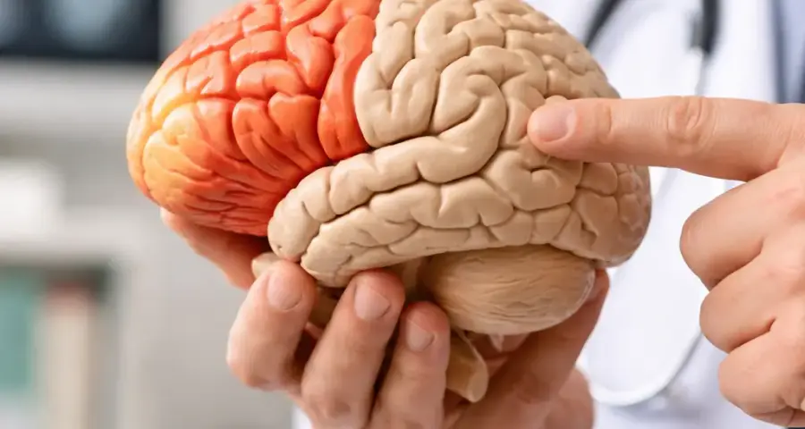
1. Cerebral Cortex: The Seat of Higher Cognition
The cerebral cortex is the wrinkled outer layer of the brain responsible for the most advanced functions: language, reasoning, planning, and sensory processing. It is divided into four lobes—frontal, parietal, temporal, and occipital—each with a specialized role. Understanding cerebral cortex functions is critical because cortical damage causes some of the most recognizable neurological deficits.
Frontal Lobe
The frontal lobe governs executive functions—decision-making, problem-solving, impulse control, and voluntary movement (via the primary motor cortex). Damage here can lead to personality changes, apathy, or difficulty planning (as seen in frontal lobe dementia). Broca’s area, located in the left frontal lobe, controls speech production; injury causes expressive aphasia (non-fluent speech).
Parietal Lobe
The parietal lobe processes touch, pressure, pain, and spatial awareness. The primary somatosensory cortex receives sensory input from the body. Lesions can produce contralateral neglect (ignoring the opposite side of the body), astereognosis (inability to identify objects by touch), or difficulty with spatial orientation.
Temporal Lobe
The temporal lobe handles auditory processing, language comprehension (Wernicke’s area), and memory formation via the hippocampus. Damage to Wernicke’s area causes fluent aphasia (garbled but fluent speech with poor comprehension). Temporal lobe epilepsy often produces auras of deja vu or olfactory hallucinations.
Occipital Lobe
The occipital lobe is the primary visual processing center. Lesions cause vision loss (cortical blindness or visual field cuts). Damage to specific visual association areas can produce agnosias (inability to recognize objects) or prosopagnosia (inability to recognize faces).
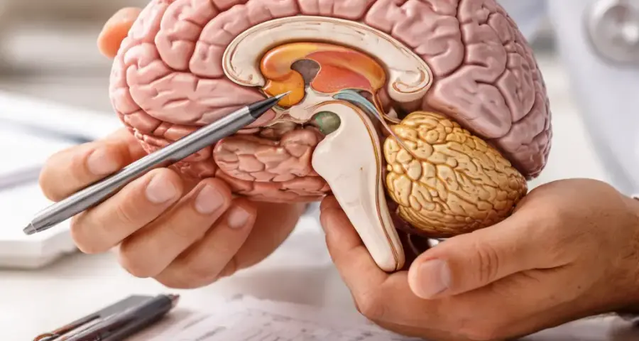
2. Thalamus: The Sensory Relay Station
The thalamus sits deep within the brain and acts as a relay and processing hub for nearly all sensory signals (except smell) before they reach the cortex. It also plays a role in motor control, arousal, and memory. Because of its central position, thalamus function is essential for normal sensory experience. Unilateral damage can cause contralateral sensory loss, ataxia, or even pain syndromes (thalamic pain syndrome or Dejerine-Roussy syndrome). Bilateral damage can lead to coma or persistent vegetative states.
Clinical correlation: In thalamic strokes, patients often present with pure sensory loss on one side of the body without weakness (lacunar syndrome).
3. Hypothalamus: The Master Regulator of Homeostasis
The hypothalamus, though small, controls body temperature, hunger, thirst, sleep-wake cycles, and endocrine function via the pituitary gland. It is also central to emotional responses (fear, anger, pleasure) and autonomic nervous system regulation. Damage can cause diabetes insipidus, hyperphagia or anorexia, temperature dysregulation, and sleep disturbances. Hypothalamic tumors or lesions are rare but devastating, often leading to obesity, endocrine deficiencies, and behavioral changes.
4. Hippocampus: The Memory Architect
The hippocampus, a seahorse-shaped structure in the medial temporal lobe, is essential for forming new declarative memories (facts and events). It also supports spatial navigation. Hippocampus function is one of the most studied topics in clinical neuroanatomy brain areas because of its role in Alzheimer’s disease, which typically begins here. Damage leads to anterograde amnesia (inability to form new memories), as famously documented in patient H.M. after bilateral temporal lobectomy. Chronic stress and depression can also shrink the hippocampus, impacting memory.
5. Amygdala: The Emotional Sentry
The amygdala, an almond-shaped nucleus in the temporal lobe, is the brain’s threat detector. It processes fear, aggression, and emotional memories. Limbic system function relies heavily on the amygdala’s connections to the hippocampus and hypothalamus. Damage can produce either excessive fear (anxiety disorders, PTSD) or reduced fear (Urbach-Wiethe disease—patients lose the ability to feel fear). In temporal lobe epilepsy, the amygdala is often involved, leading to intense emotional auras or automatisms.
6. Basal Ganglia: The Movement Coordinators
The basal ganglia are a group of deep nuclei (caudate, putamen, globus pallidus, substantia nigra, subthalamic nucleus) that fine-tune voluntary movement, prevent unwanted movements, and support habit learning. Dysfunction in basal ganglia function produces movement disorders. Parkinson’s disease (loss of dopamine in substantia nigra) causes tremor, rigidity, bradykinesia, and postural instability. Huntington’s disease (atrophy of caudate) causes chorea (involuntary dance-like movements). Dystonia and tic disorders (Tourette syndrome) also involve basal ganglia circuits.
Memory aid: The basal ganglia are like a brake and accelerator for movement—too little braking (Huntington’s) = too much movement; too much braking (Parkinson’s) = too little movement.
7. Cerebellum: The Fine-Tuner of Movement and Coordination
The cerebellum, located at the back of the brain below the occipital lobes, is responsible for motor coordination, precision, timing, and balance. It also plays a role in some cognitive functions (verbal working memory, emotional regulation). Cerebellum function is essential for smooth, coordinated movement. Damage results in ataxia (staggering gait), dysmetria (past-pointing), intention tremor, dysdiadochokinesia (difficulty with rapid alternating movements), and nystagmus. Cerebellar strokes, tumors, or degeneration (e.g., spinocerebellar ataxias) produce these classic findings.
Clinical pearl: In cerebellar disease, finger-to-nose test shows intention tremor that worsens as the finger approaches the target—a key exam finding.
8. Brainstem: The Life-Support Center
The brainstem consists of the midbrain, pons, and medulla oblongata. It houses cranial nerve nuclei (III–XII), controls breathing, heart rate, blood pressure, and arousal (reticular activating system). Brainstem roles are so vital that even small strokes here can be fatal. Locked-in syndrome (bilateral pontine damage) leaves a patient completely paralyzed but conscious, able to communicate only through eye movements. Midbrain lesions (e.g., Weber’s syndrome) produce ipsilateral oculomotor nerve palsy and contralateral hemiparesis.
Understanding brainstem cerebellum cortex functions together is crucial: the brainstem provides the basic life support and cranial nerve functions that allow the cortex and cerebellum to do their jobs.
9. Corpus Callosum: The Interhemispheric Bridge
The corpus callosum is a thick bundle of nerve fibers that connects the left and right cerebral hemispheres, allowing them to communicate. This brain region specialization is key for integrating sensory and motor information across both sides. Damage (e.g., from stroke, trauma, or callosotomy for severe epilepsy) can produce split-brain syndrome: patients cannot verbally name objects presented to their left visual field (since language is typically left-hemisphere dominant), yet can pick up the object with their left hand—demonstrating that each hemisphere processes information independently.
10. Cingulate Gyrus: The Emotional Integrator
The cingulate gyrus is a C-shaped fold of cortex above the corpus callosum. It is part of the limbic system and plays a role in emotional processing, pain perception, and decision-making (especially error detection and conflict monitoring). Anterior cingulate lesions can cause akinetic mutism (loss of willed movement and speech) or emotional blunting. Overactivity is linked to obsessive-compulsive disorder (OCD).
11. Ventricles and Cerebrospinal Fluid: The Protective Environment
The ventricular system (lateral, third, and fourth ventricles) produces and circulates cerebrospinal fluid (CSF), which cushions the brain and spinal cord, removes waste, and maintains stable pressure. Central nervous system function depends on normal CSF flow. Obstruction (e.g., from tumor, bleed, or congenital malformation) causes hydrocephalus, leading to headache, vomiting, gait disturbance, urinary incontinence, and cognitive decline (classic triad of normal pressure hydrocephalus). Ventricular anatomy is also key for CNS anatomy learning guide—knowing the location of foramina and aqueducts helps you predict where CSF will build up.
How Do Brain Structures Affect Human Behavior and Function?
The answer lies in the specialized roles described above. Each structure contributes a piece to the puzzle of human behavior. The frontal lobe governs executive function—without it, we lose the ability to plan and inhibit impulses. The hippocampus encodes memories, so without it we cannot learn from experience. The amygdala adds emotional weight—fear keeps us safe, but excessive fear can be paralyzing. The basal ganglia ensure smooth movement, and the cerebellum fine-tunes it. The brainstem keeps us alive. Understanding these neural structure function relationships is the heart of clinical neurology: when a patient comes in with a specific deficit, you can localize the lesion to a specific brain region specialization.
What Are the Clinical Roles of Major Brain Areas? (Summary Table)
| Structure | Primary Function | Clinical Correlations |
|---|---|---|
| Cerebral Cortex | Cognition, sensation, language, voluntary movement | Stroke, aphasias, neglect, dementia, epilepsy |
| Thalamus | Sensory relay, motor control, arousal | Thalamic pain syndrome, sensory stroke |
| Hypothalamus | Homeostasis, endocrine control, hunger/thirst | Diabetes insipidus, obesity, temperature dysregulation |
| Hippocampus | Memory formation, spatial navigation | Alzheimer’s disease, amnesia, depression (atrophy) |
| Amygdala | Fear, emotion, emotional memory | Phobias, PTSD, temporal lobe epilepsy, Urbach-Wiethe |
| Basal Ganglia | Motor coordination, habit learning | Parkinson’s disease, Huntington’s disease, tics, dystonia |
| Cerebellum | Motor precision, coordination, balance | Ataxia, dysmetria, intention tremor, spinocerebellar ataxias |
| Brainstem | Breathing, heart rate, cranial nerves, arousal | Locked-in syndrome, Weber’s syndrome, coma |
| Corpus Callosum | Interhemispheric communication | Split-brain syndrome, callosal stroke |
| Cingulate Gyrus | Emotion, pain, error detection | Akinetic mutism, OCD, emotional blunting |
| Ventricles / CSF | Brain cushioning, waste removal | Hydrocephalus, meningitis, CSF leaks |
How Can I Easily Memorize Brain Structures and Their Functions?
Mastering neuroanatomy study guide material requires effective strategies:
Use Mnemonics
For cranial nerves: “On Old Olympus’ Towering Top, A Finn And German Viewed Some Hops” (Olfactory, Optic, Oculomotor, Trochlear, Trigeminal, Abducens, Facial, Vestibulocochlear, Glossopharyngeal, Vagus, Spinal Accessory, Hypoglossal). For lobes: “F-P-T-O” (Frontal, Parietal, Temporal, Occipital).
Draw and Label
Sketch a lateral view of the brain and label each structure. Color-code different systems (motor = red, sensory = blue, limbic = green). Repetition builds spatial memory.
Flashcards with Clinical Scenarios
Make cards that say “Patient cannot name objects in left visual field, but can pick them up with left hand. Where is the lesion?” → Corpus callosum. This ties brain structure damage effects to real presentations.
Study in Chunks
Divide the 11 structures into three groups: Cortex (4 lobes), Deep Gray (thalamus, hypothalamus, basal ganglia, hippocampus, amygdala, cingulate), and Hindbrain/Hub (cerebellum, brainstem, corpus callosum, ventricles).
What Happens When Specific Brain Structures Are Damaged?
We have already covered each structure above, but here are a few striking examples that highlight brain structure damage effects:
- Frontal lobe damage: Personality change (like Phineas Gage—impulsive, irritable after a rod pierced his frontal lobe).
- Hippocampus damage (bilateral): Complete anterograde amnesia (patient H.M. could not form new memories after surgery).
- Amygdala damage (bilateral): Unable to recognize fear in others’ faces.
- Brainstem (pons) damage: Locked-in syndrome—complete paralysis but full awareness.
These cases are the bedrock of functional neuroanatomy clinical relevance—every deficit tells you where the lesion is.
Why Are Brain Structures Important in Neurology?
Neurology is a specialty of localization. When a patient presents with weakness on the right side of the body, a language deficit, or a visual field cut, the neurologist must determine which brain structure is affected. This is only possible with a thorough understanding of brain organization principles and brain function localization. Whether you are studying for the USMLE, preparing for rounds, or treating patients in the clinic, knowledge of Important Brain Structures and Their Clinical Functions is non-negotiable. It allows you to predict which neurological disorders and brain areas are involved, choose the right imaging study, and interpret findings accurately. For a related guide, see 9 Basic Neuroanatomy Principles You Should Never Forget.
How Do Different Parts of the Brain Communicate?
The brain is a network. Communication occurs through white matter tracts (e.g., corpus callosum, internal capsule, arcuate fasciculus) and through neurotransmitter systems (dopamine, serotonin, glutamate, GABA). Sensory information travels from the periphery → spinal cord → brainstem → thalamus → cortex. Motor commands travel from cortex → internal capsule → brainstem → spinal cord → muscle. The limbic system function requires connections between the amygdala, hippocampus, and hypothalamus. Understanding these pathways is part of neurological structure mapping and helps explain why symptoms often extend beyond a single structure. For a related guide, see Neurological Conditions: Symptoms, Signs, and Treatment.
How Are Brain Structures Linked to Neurological Diseases?
Virtually every neurological disease has a structural correlate:
- Alzheimer’s disease: Hippocampus and cortex (temporal and parietal) atrophy.
- Parkinson’s disease: Substantia nigra degeneration (basal ganglia).
- Multiple sclerosis: Demyelination in white matter tracts (can affect any structure).
- Stroke: Focal damage to one structure (e.g., middle cerebral artery stroke affects frontal, parietal, and temporal lobes).
- Epilepsy: Seizures often originate in the temporal lobe (hippocampus/amygdala).
This functional neuroanatomy clinical relevance makes studying brain structures practical and directly applicable to patient care.
Useful Resources
For deeper study of brain anatomy for medical students, explore these authoritative sources:
- Neuroanatomy Overview – NCBI Bookshelf: A free, detailed textbook covering the structure and function of all major brain regions.
- Clinical Neuroanatomy (Medical Library): A comprehensive resource for neurology trainees and clinicians.
Frequently Asked Questions About Important Brain Structures and Their Clinical Functions
What is the largest part of the brain and what does it do?
The cerebrum, which includes the cerebral cortex, is the largest part of the brain. It controls higher cognitive functions such as thinking, learning, memory, language, and voluntary movement.
Which brain structure controls balance and coordination?
The cerebellum controls balance, coordination, and fine-tuning of movement. Damage to the cerebellum leads to ataxia (uncoordinated movement) and intention tremor.
Where is the hippocampus and why is it important?
The hippocampus is located in the medial temporal lobe and is crucial for forming new declarative memories. It is one of the first areas affected in Alzheimer’s disease.
What happens if the amygdala is damaged?
Damage to the amygdala impairs the ability to recognize fear in facial expressions and reduces the startle response. It can also lead to uncharacteristic calmness or, if overactive, anxiety disorders.
What does the brainstem control?
The brainstem controls automatic life-sustaining functions including breathing, heart rate, blood pressure, and the sleep-wake cycle via the reticular activating system.
How can I remember the four lobes of the brain?
Use the mnemonic “F-P-T-O” for Frontal, Parietal, Temporal, and Occipital lobes. Alternatively, use a simple phrase like “Friendly People Talk Openly” to remember the order.
What is the clinical significance of the corpus callosum?
The corpus callosum connects the two hemispheres. Damage can lead to split-brain syndrome, where each hemisphere performs tasks independently, causing difficulties with coordinated tasks and cross-hemisphere communication.
What part of the brain is affected in Parkinson’s disease?
Parkinson’s disease involves the loss of dopamine-producing neurons in the substantia nigra, a part of the basal ganglia. This leads to tremor, rigidity, bradykinesia, and postural instability.
Which brain areas are involved in memory?
The hippocampus, amygdala, and surrounding medial temporal structures are key for memory. The prefrontal cortex helps with working memory, and the cerebellum is involved in procedural memory (motor skills).
What is the role of the thalamus?
The thalamus acts as a sensory relay station, processing and transmitting sensory information from the body to the cerebral cortex except for smell. It also contributes to motor control and arousal.
How does the hypothalamus affect behavior?
The hypothalamus regulates hunger, thirst, body temperature, and emotional responses (such as fear and anger). It also controls the endocrine system through its connection with the pituitary gland.
What is a stroke and which brain structure is most commonly affected?
Stroke is a sudden blockage or rupture of a blood vessel in the brain. The middle cerebral artery territory is most commonly affected, impacting the frontal, parietal, and temporal lobes and causing contralateral weakness and sensory loss.
What is the basal ganglia’s role in movement?
The basal ganglia fine-tune voluntary movement by inhibiting or facilitating motor signals. They prevent excessive or unwanted movements and coordinate smooth, efficient action execution.
Can brain damage affect personality?
Yes, especially damage to the frontal lobe. As seen in the famous case of Phineas Gage, frontal lobe injury can lead to impulsivity, emotional lability, and significant personality changes.
What is the difference between the left and right hemispheres?
Generally, the left hemisphere is dominant for language and analytical tasks, while the right hemisphere excels at spatial awareness, face recognition, and emotional processing. However, both sides work together via the corpus callosum.
How does the limbic system function ?
The limbic system, including the amygdala, hippocampus, hypothalamus, and cingulate gyrus, regulates emotions, memory formation, motivation, and the stress response.
What is locked-in syndrome?
Locked-in syndrome is caused by damage to the brainstem (pons). Patients are fully conscious and aware but cannot move any limb or speak—they can only communicate through eye movements.
How do brain structures communicate with each other?
Neurons send electrical signals down axons, which are bundled into white matter tracts (e.g., corpus callosum, internal capsule). Neurotransmitters (such as dopamine, serotonin, and glutamate) facilitate communication across synapses.
What happens when the cerebellum is damaged?
Cerebellar damage results in ataxia (staggering gait), dysmetria (past-pointing), intention tremor, and difficulty with rapid alternating movements. It does not cause paralysis.
How can I use this guide to study for the USMLE or board exams?
Focus on memorizing the 11 structures, their functions, and at least one classic clinical syndrome for each. Use the table in this article as a quick reference and practice with clinical vignettes that test lesion localization.