
Brain Lesions and Their Clinical Presentations Key Takeaways
Understanding brain lesions and their clinical presentations is essential for any clinician evaluating patients with neurological symptoms.
- Brain lesions and their clinical presentations vary widely depending on the type: tumors, vascular insults, trauma, infections, and inflammatory conditions each produce characteristic syndromes.
- Focal neurological deficits — such as hemiparesis, aphasia, or visual field cuts — help localize the lesion to specific brain regions like the motor cortex, Broca’s area, or optic radiations.
- Early recognition of lesion type and location using neurological examination , MRI findings, and CT imaging directly guides treatment, prognosis, and patient outcomes.
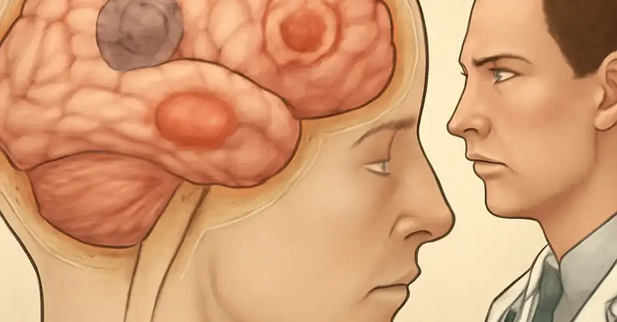
What Readers Should Know About Brain Lesions and Their Clinical Presentations
Every day in hospitals and clinics, patients present with symptoms that point to an underlying brain lesion. Whether it is a slowly growing meningioma causing chronic headaches or an acute ischemic stroke producing sudden hemiparesis, the clinical picture is shaped by three factors: where the lesion sits, how large it is, and what caused it. For medical students and neurology residents, mastering the correlation between lesion pathology and clinical findings is a cornerstone of neurological diagnosis. For a related guide, see 8 Simple Ways to Localize Neurological Lesions.
This guide walks through ten common brain lesions and their clinical presentations, from neoplasms to infectious processes, with emphasis on the neuroanatomical localization that makes each case unique. We will also discuss diagnostic tools, compare lesion types, and answer the questions most frequently asked on rotations and board exams. For a related guide, see 12 Types of Headaches and How to Differentiate Them.
The 10 Brain Lesions: Definitions, Pathophysiology, and Clinical Signs
Below are ten important lesion categories, each presented with a definition, the typical clinical presentation, underlying pathophysiology, and the most useful diagnostic approach.
1. Glioblastoma Multiforme (GBM)
Definition: GBM is a grade IV astrocytoma, the most aggressive primary brain tumor, with rapid infiltration of surrounding brain tissue.
Clinical Presentation: Patients often present with progressive headache, seizures, focal neurological deficits (e.g., hemiparesis if motor cortex is involved), and cognitive decline over weeks to months. Papilledema may be seen due to increased intracranial pressure.
Pathophysiology: GBM arises from glial cells and is characterized by neoangiogenesis, necrosis, and invasion along white matter tracts. Its rapid growth causes mass effect and edema.
Diagnostic Approach: Brain MRI findings show a ring-enhancing lesion on T1-weighted imaging with surrounding vasogenic edema on FLAIR. CT helps evaluate calcification and hemorrhage.
2. Ischemic Stroke (Cerebral Infarction)
Definition: An area of brain tissue death due to occlusion of a cerebral artery, most commonly the middle cerebral artery (MCA).
Clinical Presentation: Sudden onset of focal deficits — contralateral hemiparesis and hemisensory loss, aphasia (dominant hemisphere), neglect (non-dominant hemisphere), or homonymous hemianopia — depending on vascular territory.
Pathophysiology: Thrombotic or embolic occlusion leads to cytotoxic edema and eventual liquefactive necrosis. The penumbra of ischemic tissue around the core is salvageable if reperfusion occurs quickly.
Diagnostic Approach: CT head is used to rule out hemorrhage; MRI with diffusion-weighted imaging (DWI) shows hyperintense signal within minutes of symptom onset. Neurological examination localization matches the vascular territory.
3. Intracerebral Hemorrhage (ICH)
Definition: Bleeding directly into brain parenchyma, often due to hypertension, amyloid angiopathy, or vascular malformations.
Clinical Presentation: Abrupt severe headache, vomiting, decreased consciousness, and focal deficits that worsen over minutes to hours. Hypertension is common. Mass effect can lead to herniation.
Pathophysiology: Blood extravasates, causing direct tissue destruction and a secondary inflammatory response. Hematoma expansion can occur in the first few hours.
Diagnostic Approach: Non-contrast CT is the gold standard — hyperdense mass. MRI may show susceptibility artifact on gradient-echo sequences identifying old microbleeds.
4. Meningioma
Definition: A typically benign tumor arising from the meninges (arachnoid cap cells), often slow-growing and extra-axial.
Clinical Presentation: Seizures are the most common initial symptom. Headaches, personality changes, and focal deficits occur as the tumor compresses adjacent cortex. Many are incidental on imaging.
Pathophysiology: Meningiomas grow from the dura and compress rather than invade brain tissue. They can cause hyperostosis of the overlying skull.
Diagnostic Approach: On MRI, they appear as extra-axial, dural-based masses with homogeneous enhancement and a “dural tail.” CT shows a hyperdense lesion with possible calcification.
5. Metastatic Brain Tumors
Definition: Secondary intracranial lesions from primary cancers elsewhere, most commonly lung, breast, melanoma, renal cell, and colorectal.
Clinical Presentation: Similar to GBM — headache, seizures, and focal deficits — but often with rapid onset and multiple lesions. Systemic cancer history is key.
Pathophysiology: Hematogenous seeding to the gray-white matter junction. Vasogenic edema is prominent.
Diagnostic Approach: MRI with contrast shows multiple ring-enhancing lesions at the gray-white interface. CT may miss small metastases. Whole-body imaging helps find the primary.
6. Brain Abscess
Definition: A focal suppurative infection within the brain parenchyma, often from contiguous spread (sinusitis, otitis) or hematogenous seeding (endocarditis).
Clinical Presentation: Triad of headache, fever, and focal neurological deficit. Seizures and altered mental status are common. Symptoms evolve over days to weeks.
Pathophysiology: Necrosis, edema, and capsule formation. Responsible organisms include streptococci, staphylococci, and anaerobes.
Diagnostic Approach: MRI shows a ring-enhancing lesion with central restricted diffusion on DWI — this helps differentiate from tumor. Lumbar puncture is contraindicated due to risk of herniation.
7. Multiple Sclerosis (MS) Plaques
Definition: Focal demyelinating lesions in the white matter caused by autoimmune attack on oligodendrocytes.
Clinical Presentation: Episodic neurological deficits (optic neuritis, internuclear ophthalmoplegia, limb weakness, sensory disturbances) that are separated in time and space. Fatigue and Uhthoff phenomenon are common.
Pathophysiology: Perivenular inflammation leads to demyelination and axonal loss. Lesions are chronic and may show partial remyelination.
Diagnostic Approach: MRI shows ovoid, periventricular T2 hyperintensities — Dawson fingers — with enhancement in active plaques. CSF analysis shows oligoclonal bands.
8. Traumatic Brain Injury (TBI): Contusion and Diffuse Axonal Injury (DAI)
Definition: Contusions are focal cortical bruises; DAI is widespread axonal damage from shearing forces, often at the gray-white junction.
Clinical Presentation: Contusions cause focal deficits depending on location (e.g., frontal lobe — personality change). DAI presents with immediate loss of consciousness and prolonged coma, often without focal signs on exam.
Pathophysiology: Contusions involve hemorrhagic necrosis. DAI results from rotational acceleration-deceleration that tears axons.
Diagnostic Approach: CT is good for acute contusions (hemorrhagic). MRI (especially susceptibility-weighted imaging) is superior for DAI — shows punctate microhemorrhages. Neurological examination severity correlates with Glasgow Coma Scale score.
9. Cavernous Malformation (Cavernoma)
Definition: A vascular abnormality consisting of enlarged, thin-walled capillaries without intervening brain tissue, often with slow blood flow.
Clinical Presentation: Seizures are the most common presentation, followed by focal deficits from recurrent microhemorrhage or mass effect. Many are asymptomatic and found incidentally.
Pathophysiology: The cavernous channels are prone to leakage and thrombosis. They are low-pressure, but can cause significant hemorrhage in rare cases.
Diagnostic Approach: MRI shows a characteristic “popcorn” appearance on T2-weighted imaging with a surrounding hemosiderin rim (T2* hypointensity). CT is often normal.
10. Cerebral Toxoplasmosis
Definition: An opportunistic infectious lesion caused by Toxoplasma gondii, typically seen in immunocompromised patients (HIV/AIDS).
Clinical Presentation: Fever, headache, confusion, seizures, and focal deficits (e.g., hemiparesis). Encephalopathy is common. Multiple lesions often involve the basal ganglia and gray-white junction.
Pathophysiology: Reactivation of latent cysts leads to necrotizing encephalitis and microglial nodules. CD4 count is usually below 100 cells/µL.
Diagnostic Approach: MRI shows multiple ring-enhancing lesions with eccentric “target” appearance. Serology (IgG positive) and clinical context (HIV+ patient) are key. Biopsy may be needed in atypical cases.
| Lesion Type | Typical Location | Key Clinical Sign | Best Imaging |
|---|---|---|---|
| Glioblastoma Multiforme | White matter, frontal/temporal | Progressive headache, seizures | MRI T1 with contrast (ring enh.) |
| Ischemic Stroke | Vascular territory (MCA most common) | Sudden hemiparesis, aphasia | MRI DWI |
| Intracerebral Hemorrhage | Basal ganglia, thalamus, cerebellum | Acute headache, coma | Non-contrast CT |
| Meningioma | Supratentorial, parasagittal | Seizures, incidental | MRI T1 + gadolinium |
| Metastatic Tumor | Gray-white junction | Multiple lesions, rapid onset | MRI T1 + gadolinium |
| Brain Abscess | Frontal/temporal (sinus), any site | Fever, focal deficit, headache | MRI DWI (restricted diffusion) |
| MS Plaques | Periventricular, corpus callosum | Relapsing-remitting deficits | MRI T2/FLAIR |
| TBI Contusion | Frontal, temporal poles | Personality change, coma | CT (acute), MRI SWI |
| Cavernous Malformation | Anywhere, often supratentorial | Seizures, microhemorrhage | MRI T2* GRE |
| Toxoplasmosis | Basal ganglia, gray-white jct. | Fever, encephalopathy, HIV+ | MRI T1 + gadolinium (multiple) |
How Diagnostic Tools Help Identify Brain Lesions
The process of diagnosing a brain lesion begins with a thorough neurological examination, which localizes the lesion based on motor, sensory, cognitive, and reflex findings. For example, a patient with a right-sided hemiparesis and expressive aphasia likely has a left frontal lobe lesion affecting Broca’s area and the motor cortex. Once a lesion is suspected, imaging confirms its presence and helps characterize it.
Imaging: The Cornerstone of Diagnosis
Brain MRI findings provide the highest resolution for soft tissue. T1-weighted imaging shows anatomy; T2-weighted and FLAIR sequences highlight edema and demyelination; gadolinium enhancement indicates blood-brain barrier breakdown. Diffusion-weighted imaging (DWI) is highly sensitive for acute ischemia, while susceptibility-weighted imaging (SWI) detects microbleeds in DAI and cavernomas. Computed tomography scans are faster and excellent for detecting acute hemorrhage, mass effect, and calcifications. CT angiography can identify vascular occlusions or aneurysms.
Additional Diagnostic Tests
Lumbar puncture is used when infection or demyelination is suspected (e.g., meningitis, MS). Electroencephalography (EEG) helps evaluate seizure disorders related to cortical lesions. Biopsy remains the gold standard for definitive diagnosis of many neoplasms and infections. Advanced tools like MR spectroscopy, perfusion imaging, and functional MRI provide additional metabolic and hemodynamic information.
Comparing Lesion Types: Clinical Presentation, Progression, and Outcomes
Lesions differ not just in cause but in tempo. Vascular lesions (stroke, hemorrhage) present acutely over seconds to hours. Tumors and infections evolve over days to weeks. Inflammatory conditions like MS follow a relapsing-remitting course. Progression is also influenced by size — a 5 cm GBM in the frontal lobe may present only with subtle personality change, while a 2 cm lesion in the internal capsule can cause dense hemiparesis. Treatment approaches vary widely, from surgical resection to thrombolysis, antibiotics, steroids, or immunotherapy. Outcomes depend on pathology, location, and timeliness of intervention.
Why Early Recognition Is Critical
Recognizing the signs of a brain lesion early can be the difference between a full recovery and permanent disability. For ischemic stroke, every minute of delay loses 1.9 million neurons. For a brain abscess, timely antibiotics and drainage prevent herniation. For MS, early disease-modifying therapy reduces the number of relapses. The ability to correlate neurological examination findings with neuroanatomy and brain MRI findings is a skill every clinician must develop. This knowledge directly improves patient care and outcomes. For a related guide, see 9 Basic Neuroanatomy Principles You Should Never Forget.
Frequently Asked Questions About Brain Lesions and Their Clinical Presentations
What are the most common brain lesions and their clinical presentations ?
The most common include ischemic stroke (sudden hemiparesis, aphasia), glioblastoma (progressive headache, seizures), meningioma (often asymptomatic or seizures), intracerebral hemorrhage (acute headache, coma), and metastases (multiple lesions, rapid focal deficits).
How do brain lesions affect neurological function?
They disrupt the normal electrical and chemical activity of neurons. Depending on the location, this can produce motor weakness, sensory loss, visual deficits, language impairment, cognitive decline, or seizures. The effect is proportional to the lesion size and how critical the affected region is for that function.
What symptoms indicate a brain lesion?
Common warning signs include new-onset seizures, progressive headache (especially with morning worsening), weakness on one side of the body, difficulty speaking, vision changes, confusion, personality changes, and loss of coordination. Any abrupt or progressive neurological deficit warrants evaluation.
How are brain lesions diagnosed by doctors?
Doctors begin with a detailed history and neurological examination to localize the lesion. This is followed by imaging — typically CT for acute issues and MRI for detailed characterization. Additional tests like EEG, lumbar puncture, or biopsy may be needed based on suspected etiology.
What causes different types of brain lesions?
Causes include tumors (benign or malignant), vascular events (ischemic stroke, hemorrhage, malformations), trauma (contusion, DAI), infections (abscess, toxoplasmosis), inflammatory diseases (MS), and degenerative processes. Each has a distinct pathogenesis.
How do brain lesion symptoms vary by location?
Frontal lobe lesions can cause personality changes and motor deficits; parietal — sensory loss and neglect; temporal — memory issues and aphasia; occipital — visual field defects; cerebellum — ataxia and coordination problems; brainstem — cranial nerve palsies and long tract signs. Location is everything in clinical correlation.
What is the difference between benign and malignant brain lesions?
Benign lesions (e.g., meningioma, cavernoma) grow slowly, do not invade surrounding tissue, and can often be cured with surgery. Malignant lesions (e.g., glioblastoma, metastases) grow rapidly, infiltrate brain tissue, and carry a worse prognosis. Malignancy is defined by histology and behavior.
Can brain lesions cause cognitive and behavioral changes?
Yes, especially lesions in the frontal and temporal lobes. Patients may experience memory loss, executive dysfunction, apathy, disinhibition, depression, or psychosis. Cognitive deficits are common in large tumors, MS, and traumatic brain injury.
How do brain lesions affect movement and coordination?
Motor cortex or corticospinal tract lesions cause weakness (hemiparesis). Cerebellar lesions cause ataxia, dysmetria, and intention tremor. Basal ganglia lesions can produce movement disorders like chorea or parkinsonism. Coordination relies on intact cerebellar circuits.
What imaging tests are used to detect brain lesions?
CT is the first-line for acute hemorrhage, mass effect, and fractures. MRI with and without contrast is the gold standard for detailed anatomy, characterization, and differential diagnosis. Advanced sequences like DWI, SWI, and MR spectroscopy add specificity.
Can brain lesions lead to seizures?
Yes, seizures are a common presentation of many brain lesions, especially tumors (meningioma, GBM), cavernous malformations, contusions, and abscesses. The lesion irritates the surrounding cortex, creating a focus for abnormal electrical activity.
What treatment options are available for brain lesions?
Treatment is lesion-specific and includes surgical resection, radiation, chemotherapy, antiepileptic drugs, antibiotics, steroids, thrombolysis, endovascular procedures, or immunotherapy. Management often involves a multidisciplinary team including neurology, neurosurgery, oncology, and radiology.
How does lesion size influence clinical symptoms?
Larger lesions generally cause more symptoms and more severe deficits, but location modifies this rule. A small lesion in the internal capsule or brainstem can be devastating, while a large slow-growing frontal tumor may produce only subtle personality changes due to neural compensation.
What are the warning signs of serious brain lesions?
Acute severe headache, sudden weakness or numbness, difficulty speaking, vision loss, seizure with fever, confusion, neck stiffness, vomiting, or rapidly declining consciousness require immediate attention. Any of these can indicate a life-threatening lesion.
How do neurologists evaluate patients with brain lesions?
Neurologists perform a systematic neurological examination, including mental status, cranial nerves, motor, sensory, reflexes, coordination, and gait. They use the findings to localize the lesion, then order appropriate imaging and labs to determine the etiology and urgency.
Are all brain lesions cancerous?
No. Many brain lesions are non-cancerous, including meningiomas, cavernous malformations, MS plaques, and post-stroke cysts. Benign lesions can still cause significant symptoms if located in critical areas, but they do not metastasize.
How do vascular brain lesions differ from tumors?
Vascular lesions (stroke, hemorrhage, malformations) present acutely with deficits that are often maximal at onset. Tumors present more gradually over weeks to months. Imaging features also differ — vascular lesions follow vascular territories, while tumors have mass effect and enhancement patterns.
Can brain lesions in children present differently?
Yes. Children may present with nonspecific symptoms like vomiting, irritability, head tilt, or developmental regression. Posterior fossa tumors (e.g., medulloblastoma) are more common in children, causing hydrocephalus and ataxia. Early diagnosis relies on a high index of suspicion.
What is the prognosis for patients with brain lesions?
Prognosis is highly variable. Benign meningiomas have excellent outcomes after resection. Ischemic stroke outcomes depend on size, location, and prompt treatment. Glioblastoma has a poor prognosis (median survival ~15 months). Abscesses often resolve with antibiotics. The key factors are lesion type, size, location, and underlying patient health.
How can medical students best learn neuroanatomy clinical correlation ?
Practice with case-based learning: start with the patient’s symptom, trace the likely lesion location using neuroanatomy knowledge, then correlate with imaging. Using atlases, online resources, and cadaver dissection helps. The table in this article is a starting point for understanding typical presentations.