Cranial Nerves and Their Functions Key Takeaways
Understanding Cranial Nerves and Their Functions is essential for medical and nursing students because these twelve paired nerves control everything from smelling your morning coffee to blinking, chewing, and speaking.
- There are twelve cranial nerves numbered I through XII, each with a specific role in sensation, movement, or both.
- Mnemonics like "Oh Oh Oh To Touch And Feel Very Green Vegetables AH" help students memorize the nerve names in order.
- Clinical exams assess cranial nerve function through simple bedside tests that can detect strokes, tumors, and nerve palsies.
What Are the 12 Cranial Nerves and Their Functions? A Simplified Overview
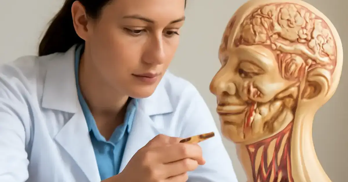
The human nervous system includes twelve pairs of cranial nerves that emerge directly from the brain (not the spinal cord). They are the communication highway between your brain and your head, neck, and torso. For healthcare trainees, mastering cranial nerve functions is non-negotiable: almost every neurological complaint—from double vision to facial droop—involves one of these nerves. For a related guide, see 12 Key Steps in the Neurological Examination Explained Simply.
Each nerve is assigned a Roman numeral (I through XII) and a name that reflects its primary role. The grouping is based on embryological origin and the type of information they carry. Understanding this organization is the first step in building your neuroanatomy basics.
Why Cranial Nerve Anatomy Matters in Clinical Practice
When you perform a neurological examination, you are essentially checking the health of each cranial nerve. A patient who cannot smell may have damage to cranial nerve I (olfactory). A patient with slurred speech might have a lesion on cranial nerve XII (hypoglossal). By connecting structure to function, you create a mental map that guides diagnosis.
Complete Guide to Each Cranial Nerve: Sensory, Motor, or Mixed
Below is a nerve-by-nerve breakdown. Remember: sensory cranial nerves carry information toward the brain, motor cranial nerves carry commands from the brain to muscles, and mixed cranial nerves do both.
Cranial Nerve I – Olfactory Nerve (Sensory)
The olfactory nerve is the only cranial nerve that can regenerate throughout life. It transmits smell information from the nasal cavity to the brain. Damage results in anosmia (loss of smell), which can be an early sign of Parkinson’s disease or COVID-19 infection. Testing is simple: have the patient identify familiar scents (coffee, mint) with eyes closed. For a related guide, see 8 Warning Signs of Multiple Sclerosis.
Cranial Nerve II – Optic Nerve (Sensory)
The optic nerve is responsible for vision. It carries signals from the retina to the occipital lobe. A fundoscopic exam allows clinicians to visualize the optic disc for signs of papilledema (swelling) or optic atrophy. Visual acuity and visual field testing are standard parts of any cranial nerve assessment.
Cranial Nerve III – Oculomotor Nerve (Motor)
The oculomotor nerve controls most eye movements: it innervates four of the six extraocular muscles, raises the upper eyelid, and constricts the pupil. A lesion causes ptosis (drooping eyelid), outward deviation of the eye (“down and out” position), and a dilated pupil. This is a neurosurgical emergency if caused by an aneurysm.
Cranial Nerve IV – Trochlear Nerve (Motor)
The trochlear nerve is the thinnest cranial nerve and the only one that emerges from the dorsal side of the brainstem. It controls the superior oblique muscle, which rotates the eye downward and outward. Damage causes vertical diplopia (double vision), especially when looking down (e.g., reading stairs).
Cranial Nerve V – Trigeminal Nerve (Mixed)
The trigeminal nerve is the largest cranial nerve. Its three divisions (ophthalmic, maxillary, mandibular) provide sensation to the face, scalp, and cornea, and its motor branch controls the muscles of mastication (chewing). Trigeminal neuralgia—a condition of severe facial pain—is a classic disorder affecting this nerve. The corneal reflex tests the sensory (afferent) and motor (efferent) limbs of cranial nerves V and VII.
Cranial Nerve VI – Abducens Nerve (Motor)
The abducens nerve innervates the lateral rectus muscle, which abducts the eye (pulls it outward). This nerve is often the first to be affected by increased intracranial pressure, causing a convergent squint. Testing involves having the patient follow a target to the side.
Cranial Nerve VII – Facial Nerve (Mixed)
The facial nerve controls the muscles of facial expression, taste from the anterior two-thirds of the tongue, and parasympathetic innervation to the salivary and lacrimal glands. Bell’s palsy (a sudden peripheral facial paralysis) is the most common disorder. Testing includes asking the patient to smile, puff cheeks, and raise eyebrows.
Cranial Nerve VIII – Vestibulocochlear Nerve (Sensory)
This nerve has two components: the cochlear branch (hearing) and the vestibular branch (balance). Hearing loss, tinnitus, and vertigo are common complaints. The Rinne and Weber tuning fork tests are simple bedside tools to differentiate conductive from sensorineural hearing loss. Caloric testing assesses vestibular function.
Cranial Nerve IX – Glossopharyngeal Nerve (Mixed)
The glossopharyngeal nerve provides taste to the posterior one-third of the tongue, sensation to the pharynx and tonsils, and motor control to the stylopharyngeus muscle (involved in swallowing). The gag reflex tests cranial nerves IX and X together. Lesions cause dysphagia and loss of taste in the posterior tongue.
Cranial Nerve X – Vagus Nerve (Mixed)
The vagus nerve is the longest cranial nerve, extending into the thorax and abdomen. It controls the muscles of the pharynx and larynx (voice production), provides parasympathetic innervation to the heart and digestive tract, and carries sensory information from the ear and larynx. Hoarseness or a weak cough suggests vagal damage. The “say ahh” part of an exam tests the vagus nerve’s motor function.
Cranial Nerve XI – Accessory Nerve (Motor)
The spinal accessory nerve innervates the trapezius and sternocleidomastoid muscles. Testing involves shoulder shrug (trapezius) and head turn against resistance (sternocleidomastoid). Weakness on one side can indicate a lesion. This nerve is sometimes injured during neck surgeries.
Cranial Nerve XII – Hypoglossal Nerve (Motor)
The hypoglossal nerve controls all muscles of the tongue except the palatoglossus. When asked to protrude the tongue, a patient with hypoglossal damage will deviate toward the weak side. Fasciculations (twitching) of the tongue are a classic sign of lower motor neuron disease such as amyotrophic lateral sclerosis (ALS).
| Nerve | Name | Type | Primary Function |
|---|---|---|---|
| I | Olfactory | Sensory | Smell |
| II | Optic | Sensory | Vision |
| III | Oculomotor | Motor | Eye movement, pupil constriction |
| IV | Trochlear | Motor | Eye movement (down and inward) |
| V | Trigeminal | Mixed | Facial sensation, chewing |
| VI | Abducens | Motor | Eye movement (lateral) |
| VII | Facial | Mixed | Facial expression, taste (anterior 2/3) |
| VIII | Vestibulocochlear | Sensory | Hearing, balance |
| IX | Glossopharyngeal | Mixed | Taste (posterior 1/3), swallowing |
| X | Vagus | Mixed | Swallowing, voice, parasympathetic control |
| XI | Accessory | Motor | Neck and shoulder movement |
| XII | Hypoglossal | Motor | Tongue movement |
How Cranial Nerves Contribute to Vision, Hearing, Taste, Smell, Facial Expression, Swallowing, Speech, and Eye Movements
The magic of daily life—seeing a friend’s face, tasting your lunch, speaking a sentence—depends on the seamless cooperation of multiple cranial nerves. Let’s connect each function to the nerves responsible.
Vision and Eye Movements
Vision starts with the optic nerve (CN II) transmitting visual data. Coordinated eye movements require cranial nerves III, IV, and VI working together. The oculomotor nerve moves the eye up, down, and inward; the trochlear nerve rotates the eye down and outward; and the abducens nerve pulls the eye laterally. Damage to any one of these motor cranial nerves causes double vision or misalignment.
Hearing and Balance
The vestibulocochlear nerve (CN VIII) handles both hearing and balance. The cochlear division processes sound waves; the vestibular division senses head position and motion. Cranial nerve disorders affecting this nerve include acoustic neuroma, which causes unilateral hearing loss and tinnitus.
Taste and Smell
Smell is mediated by the olfactory nerve (CN I). Taste involves three nerves: the facial nerve (CN VII) carries taste from the anterior tongue, the glossopharyngeal nerve (CN IX) from the posterior tongue, and the vagus nerve (CN X) from the epiglottis and pharynx. Together, they create the flavor experience.
Facial Expression and Swallowing
The facial nerve (CN VII) controls all muscles of facial expression—smiling, frowning, raising eyebrows. Swallowing requires coordination between the trigeminal (CN V), facial (CN VII), glossopharyngeal (CN IX), vagus (CN X), and hypoglossal (CN XII) nerves. Swallowing problems (dysphagia) often involve damage to multiple lower cranial nerves.
Speech
Speech production relies on the vagus nerve (CN X) for laryngeal muscle control (voice), the facial nerve (CN VII) for lip movement, and the hypoglossal nerve (CN XII) for tongue articulation. A stroke affecting the hypoglossal nerve causes slurred speech (dysarthria).
Neurological Examination: How Clinicians Assess Cranial Nerve Function
A cranial nerve assessment is a routine part of every neurological exam. It follows a systematic order, often starting with CN I and ending with CN XII. Here is the standard approach:
- CN I (Olfactory): Test each nostril separately with a familiar scent.
- CN II (Optic): Test visual acuity with a Snellen chart and visual fields by confrontation.
- CN III, IV, VI (Oculomotor, Trochlear, Abducens): Assess extraocular movements in six cardinal gaze directions; check pupil light reflex.
- CN V (Trigeminal): Test facial sensation to light touch and pinprick in all three divisions; assess jaw jerk and masseter muscle strength.
- CN VII (Facial): Observe symmetry at rest; ask the patient to smile, puff cheeks, and close eyes tightly.
- CN VIII (Vestibulocochlear): Perform Rinne and Weber tests with a tuning fork; check for nystagmus.
- CN IX, X (Glossopharyngeal, Vagus): Assess gag reflex; ask the patient to say “ahh” and observe palate elevation; check voice quality.
- CN XI (Accessory): Test shoulder shrug and head rotation against resistance.
- CN XII (Hypoglossal): Ask the patient to protrude the tongue; look for deviation and fasciculations.
Localizing a lesion to a specific nerve is one of the most rewarding skills in clinical neurology. For example, a patient with a stroke may present with contralateral weakness but no cranial nerve deficits, while a patient with a brainstem tumor may have multiple cranial nerve palsies.
Comparison of Cranial Nerves: Anatomical Pathways, Functions, and Clinical Significance
When studying cranial nerve anatomy, it helps to compare nerves by their origin, course, and target structures. Here are key comparisons:
- Pure sensory vs. mixed: CN I, II, and VIII are sensory cranial nerves. CN V, VII, IX, and X are mixed cranial nerves. The rest are motor cranial nerves.
- Intracranial vs. extracranial course: CN III, IV, and VI travel through the cavernous sinus; CN V, VII, and VIII pass through the internal acoustic meatus.
- Clinical red flags: An isolated CN III palsy with pupil dilation is an emergency (aneurysm). Bilateral facial weakness suggests Guillain-Barré syndrome. Nystagmus + vertigo points to CN VIII or brainstem involvement.
Memory Techniques and Mnemonics for Cranial Nerves
Using cranial nerve mnemonics is the fastest way to commit the order and classification to memory. Here are three proven strategies:
Classic Mnemonic for Nerve Names (Order)
“Oh Oh Oh To Touch And Feel Very Green Vegetables AH” corresponds to: Olfactory, Optic, Oculomotor, Trochlear, Trigeminal, Abducens, Facial, Vestibulocochlear, Glossopharyngeal, Vagus, Accessory, Hypoglossal.
Mnemonic for Sensory, Motor, or Mixed
“Some Say Marry Money But My Brother Says Big Brains Matter More” – S (Sensory), S (Sensory), M (Motor), M (Motor), M (Mixed), M (Motor), M (Mixed), S (Sensory), M (Mixed), M (Mixed), M (Motor), M (Motor).
Visual and Spaced-Repetition Techniques
Draw a simple brainstem cross-section and label where each nerve emerges. Use spaced-repetition apps like Anki with flashcards that ask “What is the function of CN VII?” and “Which cranial nerves are sensory?” These anatomy study guide methods reduce cramming and improve long-term retention for anyone in medical education.
Common Cranial Nerve Disorders and What to Look For
Cranial nerve disorders manifest in predictable ways. Recognizing the pattern helps narrow the differential:
- Olfactory groove meningioma: Unilateral anosmia (CN I).
- Pituitary tumor: Bitemporal hemianopia due to optic chiasm compression (CN II).
- Third nerve palsy: Ptosis, mydriasis, eye turns “down and out”.
- Trigeminal neuralgia: Severe lancinating facial pain (CN V).
- Bell’s palsy: Acute peripheral facial paralysis (CN VII).
- Acoustic neuroma: Unilateral hearing loss and tinnitus (CN VIII).
- Bulbar palsy: Dysphagia, dysarthria, fasciculating tongue (CN IX, X, XII).
Learning these patterns is a core part of clinical neurology and will serve you well in exams and bedside practice.
Conclusion: Cranial Nerves and Their Functions Simplified for Lasting Mastery
Understanding Cranial Nerves and Their Functions doesn’t have to be overwhelming. By breaking them down into sensory, motor, and mixed categories, using mnemonics to lock in the order, and connecting each nerve to a real clinical test, you build a reliable mental framework that supports every future patient encounter. Keep this anatomy study guide nearby, practice the exam steps with a partner, and revisit the table before any neurology rotation or exam. For a related guide, see 11 Important Brain Structures and Their Clinical Functions.
Useful Resources
For further practice, explore these trusted resources:
- NCBI Bookshelf: Neuroanatomy, Cranial Nerves – detailed anatomy and clinical correlations
- TeachMeAnatomy: Cranial Nerves – visual guides and interactive quizzes
Frequently Asked Questions About Cranial Nerves and Their Functions
What are the 12 cranial nerves and their functions?
The 12 cranial nerves are: Olfactory (smell), Optic (vision), Oculomotor (eye movement, pupil constriction), Trochlear (eye movement down/inward), Trigeminal (facial sensation, chewing), Abducens (lateral eye movement), Facial (facial expression, taste), Vestibulocochlear (hearing, balance), Glossopharyngeal (taste, swallowing), Vagus (voice, swallowing, parasympathetic), Accessory (shoulder/neck movement), and Hypoglossal (tongue movement).
How can you memorize the cranial nerves easily?
Use the classic mnemonic “Oh Oh Oh To Touch And Feel Very Green Vegetables AH” for the names in order. For sensory/motor classification, use “Some Say Marry Money But My Brother Says Big Brains Matter More.” Combine with spaced-repetition flashcards and anatomy drawings for best results.
What is the function of each cranial nerve?
Each nerve has a distinct role: CN I – smell; CN II – vision; CN III – eye movement and pupil control; CN IV – eye movement; CN V – facial sensation and chewing; CN VI – lateral eye movement; CN VII – facial expressions and taste; CN VIII – hearing and balance; CN IX – taste and swallowing; CN X – voice, swallowing, internal organ regulation; CN XI – neck and shoulder movement; CN XII – tongue movement.
Which cranial nerves are sensory motor or mixed?
Sensory cranial nerves are I (Olfactory), II (Optic), and VIII (Vestibulocochlear). Motor cranial nerves are III, IV, VI, XI, and XII. Mixed cranial nerves (carrying both sensory and motor fibers) are V (Trigeminal), VII (Facial), IX (Glossopharyngeal), and X (Vagus).
How do cranial nerves affect vision hearing and smell?
Vision is mediated by the optic nerve (CN II) and coordinated by CN III, IV, and VI for eye movement. Hearing is processed by the vestibulocochlear nerve (CN VIII). Smell is transmitted by the olfactory nerve (CN I). Damage to any of these nerves impairs the corresponding sense.
What symptoms occur when cranial nerves are damaged?
Symptoms vary by nerve: loss of smell (CN I), vision loss (CN II), double vision or drooping eyelid (CN III, IV, VI), facial pain or numbness (CN V), facial droop (CN VII), hearing loss or vertigo (CN VIII), difficulty swallowing (CN IX, X), hoarseness (CN X), shoulder weakness (CN XI), or tongue deviation (CN XII).
How are cranial nerves tested during neurological exams?
Testing is systematic: smell with familiar scents (CN I), visual acuity and fields (CN II), extraocular movements and pupil reflex (CN III, IV, VI), facial sensation and jaw strength (CN V), facial symmetry (CN VII), hearing tests and balance (CN VIII), gag reflex and voice (CN IX, X), shoulder shrug (CN XI), and tongue protrusion (CN XII).
Why are cranial nerves important in human anatomy?
Cranial nerves are vital because they connect the brain directly to the sense organs, muscles of the face and neck, and internal organs. They control critical functions such as vision, hearing, balance, taste, swallowing, speech, and facial expression. Damage to cranial nerves can severely impact quality of life.
What are common disorders involving cranial nerves?
Common disorders include olfactory groove meningioma (CN I), optic neuritis (CN II), third nerve palsy (CN III), trigeminal neuralgia (CN V), Bell’s palsy (CN VII), acoustic neuroma (CN VIII), glossopharyngeal neuralgia (CN IX), and hypoglossal nerve palsy (CN XII).
How do doctors assess cranial nerve function?
Doctors perform a structured cranial nerve exam that includes testing smell, vision, eye movements, facial sensation, hearing, gag reflex, shoulder strength, and tongue movement. This assessment is a core part of the neurological exam and helps localize brainstem or peripheral nerve lesions.
What mnemonics help students remember cranial nerves?
Popular mnemonics include “Oh Oh Oh To Touch And Feel Very Green Vegetables AH” for nerve names and “Some Say Marry Money But My Brother Says Big Brains Matter More” for sensory/motor classification. Many students also create their own visual or story-based mnemonics.
How do cranial nerves connect the brain to the body?
Cranial nerves emerge directly from the brain (specifically the cerebrum, midbrain, pons, and medulla) and extend through foramina in the skull to reach their target organs. Unlike spinal nerves, they do not pass through the spinal cord, giving them a distinct role in head and neck function.
What clinical signs indicate cranial nerve injury?
Signs include anosmia (CN I), visual field defects (CN II), ptosis and dilated pupil (CN III), vertical diplopia (CN IV), diminished facial sensation (CN V), lateral gaze palsy (CN VI), facial asymmetry (CN VII), hearing loss (CN VIII), absent gag reflex (CN IX, X), hoarseness (CN X), shoulder drop (CN XI), and tongue deviation (CN XII).
How do cranial nerves support daily activities?
Everyday tasks like reading, eating, talking, smiling, and walking rely on intact cranial nerves. The optic nerve helps you see a menu, the trigeminal and facial nerves allow you to chew and taste, the vagus and hypoglossal nerves control swallowing and speech, and the vestibular nerve helps you maintain balance.
What should medical students know about cranial nerve anatomy ?
Students should memorize the 12 nerves in order, their sensory/motor/mixed classification, their brainstem origin, the foramina they exit, and their primary functions. Clinical correlations—such as which cranial nerves are affected by a cavernous sinus syndrome or acoustic neuroma—are essential for board exams and patient care.
Can cranial nerves be affected by stroke?
Yes. A brainstem stroke can damage cranial nerve nuclei, causing ipsilateral CN deficits and contralateral limb weakness (alternating syndromes). A hemispheric stroke usually spares cranial nerves, except for a contralateral lower facial weakness (CN VII) due to corticobulbar tract involvement.
What is the difference between upper and lower motor neuron lesions of cranial nerves?
Upper motor neuron lesions (e.g., stroke) cause contralateral weakness of the lower face only (CN VII) and spastic tongue (CN XII). Lower motor neuron lesions (e.g., Bell’s palsy) cause ipsilateral weakness of the entire face and flaccid, fasciculating tongue.
Which cranial nerves are most commonly injured in head trauma?
The olfactory nerve (CN I) is most vulnerable due to its location in the cribriform plate. The optic nerve (CN II), oculomotor nerve (CN III), and facial nerve (CN VII) are also commonly injured in skull base fractures.
How do cranial nerves differ from spinal nerves?
Cranial nerves emerge directly from the brain and are primarily responsible for head and neck function. Spinal nerves emerge from the spinal cord and innervate the trunk and limbs. Most cranial nerves are special sense or branchial arch derivatives, while spinal nerves are segmental and follow a consistent dorsal/ventral root pattern.
What is the best way to study cranial nerve anatomy for exams?
Combine a clear reference table, a mnemonic, and clinical flashcards. Watch dissection videos or use 3D anatomy apps. Practice the cranial nerve exam on a friend. The more you link structure to a real patient scenario, the more naturally you will recall the information under pressure.