Neurological Reflexes and What They Mean Clinically Key Takeaways
Reflex testing is a cornerstone of the neurological examination because it provides objective, reproducible information about the functional integrity of sensory and motor pathways.
- Neurological reflexes and what they mean clinically are assessed using a standardized reflex grading system —from 0 (absent) to 4+ ( hyperreflexia with clonus )—to detect subtle abnormalities.
- Deep tendon reflexes such as the biceps, triceps, patellar, and Achilles reflexes are the most commonly tested; asymmetry or a change in amplitude often points to a specific spinal segment or peripheral nerve lesion.
- Pathological reflexes like the Babinski sign and clonus are always abnormal in adults and indicate disruption of upper motor neuron pathways, helping localize damage to the corticospinal tract.
Understanding the Role of Neurological Reflexes in Clinical Diagnosis
Reflexes are involuntary, stereotyped motor responses elicited by a specific sensory stimulus. The clinical examination of neurological reflexes and what they mean clinically allows the examiner to test the integrity of the reflex arc—a simple circuit involving a sensory neuron, a synapse in the spinal cord or brainstem, and a motor neuron projecting to a muscle. When this arc is intact, a brisk and predictable muscle contraction occurs. When the arc is disrupted at any point, the reflex becomes diminished or absent. Conversely, when descending inhibitory pathways from the brain are damaged, reflexes become exaggerated. This simple principle forms the basis for localizing neurological disease. For a related guide, see 12 Key Steps in the Neurological Examination Explained Simply.
The Reflex Arc: Anatomy and Physiology
The monosynaptic stretch reflex arc begins when a muscle spindle detects stretch. The sensory (afferent) fiber enters the dorsal root, synapses directly onto an alpha motor neuron in the ventral horn, and the motor (efferent) fiber causes the muscle to contract. This basic circuit is modulated by descending tracts from the brain, which normally exert an inhibitory influence. Understanding this neuroanatomy reflex pathway is essential because a lesion anywhere along the arc—peripheral nerve, dorsal root, spinal cord gray matter, or ventral root—will reduce or abolish the reflex, while a lesion in the descending pathways will release the reflex from inhibition, producing hyperreflexia.
The Reflex Grading System: Quantifying the Neurological Examination
Before interpreting individual reflexes, clinicians must master the reflex grading system. The most widely used scale is the Mayo Clinic 0–4+ scale:
| Grade | Description | Clinical Significance |
|---|---|---|
| 0 | No response | Indicates complete disruption of the reflex arc (lower motor neuron or peripheral nerve lesion) |
| 1+ | Trace or diminished response | Suggests partial damage to the reflex arc or mild neuropathy |
| 2+ | Normal, active response | Expected in healthy individuals |
| 3+ | Brisk, more active than usual | May be normal in anxious patients; can indicate mild upper motor neuron lesion |
| 4+ | Very brisk with clonus | Strongly indicates an upper motor neuron lesion |
Grading is always performed with the patient relaxed and the limb positioned to optimize the stretch. Asymmetry—a difference of one grade or more between sides—is often more clinically significant than the absolute grade.
10 Neurological Reflexes and Their Clinical Significance
Below are the ten most clinically relevant reflexes, organized by anatomical region, with explanations of what each finding means in practice.
1. Biceps Reflex (C5–C6)
Technique: The examiner places a thumb over the biceps tendon in the antecubital fossa and strikes the thumb with the reflex hammer. A normal response is flexion of the elbow.
Clinical meaning: A diminished or absent biceps reflex suggests a lesion of the C5 or C6 nerve roots (e.g., cervical radiculopathy) or the musculocutaneous nerve. Hyperreflexia of the biceps reflex often accompanies upper motor neuron lesions above the C5 level, such as in cervical myelopathy.
2. Triceps Reflex (C7–C8)
Technique: With the patient’s arm passively flexed at the elbow, the examiner taps the triceps tendon just above the olecranon. A normal response is extension of the elbow.
Clinical meaning: An absent triceps reflex commonly occurs in C7 radiculopathy. A hyperactive triceps reflex, especially when combined with other upper motor neuron signs, points to a lesion of the corticospinal tract above C7.
3. Brachioradialis Reflex (C5–C6)
Technique: Tap the styloid process of the radius with the forearm in a neutral position. The normal response is flexion and supination of the forearm.
Clinical meaning: This reflex is often assessed together with the biceps reflex. An inverted brachioradialis reflex—where tapping the radius produces finger flexion instead of forearm flexion—is a classic sign of a C5–C6 lesion with accompanying upper motor neuron pathology below the level of the lesion (e.g., in syringomyelia).
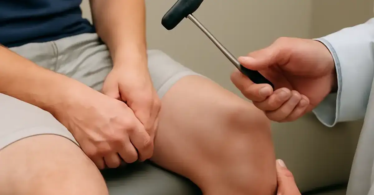
4. Patellar Reflex (L3–L4)
Technique: With the patient seated and legs hanging freely, tap the patellar tendon just below the kneecap. The normal response is extension of the knee.
Clinical meaning: The patellar reflex is one of the most commonly tested deep tendon reflexes. An absent patellar reflex is a hallmark of L3 or L4 radiculopathy, femoral neuropathy, or a lesion of the lumbar plexus. Hyperreflexia of the patellar reflex is seen in upper motor neuron disorders such as multiple sclerosis, spinal cord injury, or stroke.
5. Achilles Reflex (S1–S2)
Technique: With the patient kneeling in a chair or lying prone with the foot dorsiflexed, tap the Achilles tendon. The normal response is plantar flexion of the foot.
Clinical meaning: The Achilles reflex is often the first reflex to be lost in peripheral neuropathy (e.g., diabetic neuropathy). An absent Achilles reflex is also a classic sign of S1 radiculopathy (sciatica). A hyperactive Achilles reflex can occur in upper motor neuron lesions but is less reliable than the patellar reflex for this purpose.
6. Jaw Jerk Reflex (Trigeminal Nerve, Pontine Level)
Technique: With the patient’s mouth slightly open and relaxed, place a finger on the chin and tap it. The normal response is a slight closure of the jaw.
Clinical meaning: The jaw jerk is a stretch reflex of the masseter muscle. A brisk, exaggerated jaw jerk is a sign of a bilateral upper motor neuron lesion above the pons, often seen in pseudobulbar palsy or progressive supranuclear palsy. Unlike the limb reflexes, the jaw jerk is not normally present in healthy adults and is therefore always considered abnormal when easily elicited.
7. Hoffman Sign (C8–T1)
Technique: Support the patient’s hand and flick the distal phalanx of the middle finger. A positive response is adduction of the thumb (thumb flexion) and flexion of the index finger.
Clinical meaning: The Hoffman sign is a pathological reflex that indicates upper motor neuron dysfunction, typically from cervical cord compression or other corticospinal tract lesions. It is analogous to the Babinski sign in the upper extremity. A positive Hoffman sign in a patient with neck pain and hyperreflexia is highly suggestive of cervical myelopathy.
8. Babinski Reflex (Plantar Reflex, L5–S1)
Technique: Using a blunt object (e.g., the handle of the reflex hammer), stroke the lateral aspect of the sole from the heel to the ball of the foot and then medially across the metatarsal heads. The normal response in adults is flexion of all toes (plantar flexion).
Clinical meaning: The Babinski reflex is arguably the most important pathological reflex in clinical neurology. Extension of the great toe with fanning of the other toes is an abnormal response that indicates an upper motor neuron lesion affecting the corticospinal tract. In infants, the Babinski sign is normal until about 12 months of age due to incomplete myelination of the pyramidal tracts. An extensor plantar response in an adult is always abnormal and localizes the lesion to the contralateral motor cortex or corticospinal tract.
9. Chaddock Reflex (Plantar Variant)
Technique: Stroke the lateral aspect of the foot below the lateral malleolus. A positive response is the same as the Babinski reflex—great toe extension with toe fanning.
Clinical meaning: The Chaddock reflex is more sensitive than the Babinski sign in some patients, particularly when the sole is hypersensitive or the patient withdraws. It tests the same corticospinal pathway and carries the same clinical significance as the Babinski reflex.
10. Clonus (Lower Extremity)
Technique: With the patient supine, passively dorsiflex the foot at the ankle and maintain a steady stretch on the calf muscles. Clonus is defined as rhythmic, involuntary contractions of the gastrocnemius-soleus complex.
Clinical meaning: Sustained clonus (5 beats or more) is a sign of upper motor neuron disease. It occurs when the loss of descending inhibition leads to repetitive firing of the stretch reflex arc. Unsustained clonus (2–4 beats) can sometimes be normal, especially in anxious patients, but sustained clonus is always pathological. Clonus can also be elicited at the patella (patellar clonus) by quickly pushing the patella downward. The presence of clonus, together with hyperreflexia and the Babinski sign, constitutes the classic triad of an upper motor neuron syndrome.
Interpreting Reflex Patterns: Upper Motor Neuron vs. Lower Motor Neuron Disorders
The clinical utility of reflex examination lies in its ability to differentiate between upper motor neuron signs and lower motor neuron signs. This distinction is fundamental to the neurological diagnosis and determines the direction of further investigation.
Upper Motor Neuron (UMN) Pattern
Findings: Hyperreflexia (3+ to 4+), clonus, Babinski sign (extensor plantar response), Hoffman sign, and loss of superficial reflexes (e.g., abdominal reflexes).
Localization: Lesions in the motor cortex, internal capsule, brainstem, or spinal cord above the level of the anterior horn cell. Common causes include stroke, multiple sclerosis, spinal cord compression, and traumatic brain injury.
Lower Motor Neuron (LMN) Pattern
Findings: Hyporeflexia or areflexia (0 to 1+), absent Babinski sign (flexor plantar response), muscle atrophy, fasciculations, and flaccid weakness.
Localization: Lesions of the anterior horn cell (e.g., ALS), nerve root (radiculopathy), plexus, or peripheral nerve. Common causes include Guillain-Barré syndrome, diabetic neuropathy, and cervical or lumbar radiculopathy.
Comparison of UMN and LMN Reflex Findings
| Feature | Upper Motor Neuron | Lower Motor Neuron |
|---|---|---|
| Reflex amplitude | Hyperreflexia (3+ to 4+) | Hyporeflexia (0 to 1+) |
| Clonus | Present (sustained) | Absent |
| Babinski sign | Extensor (abnormal) | Flexor (normal) |
| Muscle tone | Spasticity | Flaccidity |
| Atrophy | Late and disuse-related | Early and severe |
| Fasciculations | Absent | Common |
How Reflex Patterns Help Localize Neurological Damage
Reflexes are organized segmentally. By testing reflexes at different spinal levels, the clinician can pinpoint the level of a spinal reflexes abnormality. For example, a patient with a C6–C7 disc herniation may have a diminished triceps reflex (C7) but a normal biceps reflex (C5–C6). A patient with a spinal cord injury at T10 will have normal upper limb reflexes but hyperreflexia and clonus in the lower limbs, with a Babinski sign bilaterally.
In peripheral neuropathies, reflex loss follows a distal-to-proximal gradient: the Achilles reflex is lost first, followed by the patellar reflex, and finally the upper limb reflexes. This pattern is highly characteristic of length-dependent axonal neuropathies such as those caused by diabetes or alcohol.
The clinical neurology exam also incorporates cranial nerve reflexes such as the pupillary light reflex (CN II and III), the corneal reflex (CN V and VII), and the gag reflex (CN IX and X). An abnormal cranial nerve reflex can localize a lesion to the brainstem or specific cranial nerve nuclei—critical information when evaluating brainstem strokes or posterior fossa tumors. For a related guide, see 10 Brain Lesions and Their Clinical Presentations Explained.
Useful Resources
For further reading on the neuroanatomy of reflex pathways and their clinical application, the following resources are authoritative and freely accessible:
- National Institute of Neurological Disorders and Stroke – Reflex Examination – Comprehensive overview of reflex testing and its role in neurological diagnosis.
- PubMed – The Utility of Deep Tendon Reflexes in Neurological Diagnosis – A review article discussing the evidence base for reflex testing in clinical practice.
Frequently Asked Questions About Neurological Reflexes and What They Mean Clinically
What are the main neurological reflexes and what do they mean clinically?
The main neurological reflexes include the biceps, triceps, brachioradialis, patellar, and Achilles deep tendon reflexes, along with pathological reflexes such as the Babinski sign and Hoffman sign. Clinically, they assess the integrity of specific spinal segments and descending motor pathways. Normal reflexes indicate an intact reflex arc, while abnormal reflexes point to nerve root, spinal cord, or brain pathology.
How do doctors test deep tendon reflexes ?
Doctors use a reflex hammer to tap a tendon, which stretches the muscle spindle and elicits a contraction. The patient must be relaxed, and the limb should be positioned to optimize the stretch. The response is graded using a standardized 0–4+ scale. Common tests include tapping the patellar tendon for the knee jerk and the Achilles tendon for the ankle jerk.
What does hyperreflexia indicate?
Hyperreflexia indicates an upper motor neuron lesion. It occurs when descending inhibitory pathways from the brain to the spinal cord are damaged, releasing the reflex arc from inhibition. Causes include stroke, multiple sclerosis, spinal cord injury, and cerebral palsy.
What causes hyporeflexia or absent reflexes?
Hyporeflexia or areflexia results from a lesion anywhere along the reflex arc—peripheral nerve, dorsal root, ventral root, or anterior horn cell. Common causes include diabetic or alcoholic neuropathy, Guillain-Barré syndrome, radiculopathy, and motor neuron disease such as ALS.
What are pathological reflexes in neurology?
Pathological reflexes are abnormal responses that are not present in healthy adults. They include the Babinski sign (extensor plantar response), Hoffman sign, clonus, and the grasp reflex. Their presence indicates a lesion of the upper motor neuron pathways, typically the corticospinal tract.
What is the Babinski reflex and what does it mean?
The Babinski reflex is elicited by stroking the lateral sole; a normal response is toe flexion. An abnormal (extensor) response—great toe extension with fanning of the other toes—indicates an upper motor neuron lesion affecting the corticospinal tract. It is one of the most reliable signs in clinical neurology.
How do reflexes help localize neurological lesions?
Each reflex is mediated by a specific spinal segment. By assessing which reflexes are abnormal, the clinician can identify the level of the lesion. For example, a diminished triceps reflex points to C7, while a diminished patellar reflex points to L3–L4. Asymmetric reflexes help lateralize the lesion to one side of the nervous system.
What is the difference between upper and lower motor neuron reflex findings?
Upper motor neuron lesions cause hyperreflexia, clonus, and a Babinski sign. Lower motor neuron lesions cause hyporeflexia or areflexia, muscle atrophy, fasciculations, and a normal plantar response. This distinction is fundamental for localizing the lesion along the neuraxis.
What are common reflex tests in a neurological exam?
The standard reflex testing battery includes the biceps (C5–C6), triceps (C7–C8), brachioradialis (C5–C6), patellar (L3–L4), and Achilles (S1–S2) reflexes. In addition, the plantar reflex (Babinski), Hoffman sign, and clonus are tested when upper motor neuron disease is suspected.
How do spinal cord lesions affect reflexes?
Spinal cord lesions produce hyperreflexia and clonus below the level of the lesion, along with a Babinski sign. At the level of the lesion, reflexes may be diminished or absent due to damage to the anterior horn cells or nerve roots. This combination of upper motor neuron findings below and lower motor neuron findings at the level helps localize the lesion.
What does clonus indicate clinically?
Sustained clonus (5 beats or more) indicates an upper motor neuron lesion. It is caused by the loss of descending inhibition, leading to repetitive, rhythmic contractions of a muscle group in response to sustained stretch. Clonus is commonly tested at the ankle and patella.
How do reflex changes help diagnose neurological diseases?
Reflex changes provide objective evidence of nervous system dysfunction. A pattern of hyperreflexia and Babinski sign suggests multiple sclerosis, stroke, or spinal cord compression. Hyporeflexia with normal sensation suggests peripheral neuropathy or radiculopathy. Reflex testing is often the first clue that guides further imaging and electrodiagnostic studies.
What are cranial nerve reflexes and their significance?
Cranial nerve reflexes are mediated by brainstem circuits and involve cranial nerves. Examples include the pupillary light reflex (CN II and III), corneal reflex (CN V and VII), and gag reflex (CN IX and X). Abnormalities help localize lesions to specific brainstem levels and are critical in assessing brainstem function, especially in coma or suspected brainstem stroke.
How are reflexes graded in clinical exams?
Reflexes are graded using a 0 to 4+ scale: 0 = absent, 1+ = diminished, 2+ = normal, 3+ = brisk, and 4+ = very brisk with clonus. The scale is subjective, but asymmetry is highly significant. Some examiners also use a 5-point scale that adds a grade for sustained clonus.
What conditions affect neurological reflex responses?
Many conditions alter reflexes: stroke, multiple sclerosis, spinal cord injury, peripheral neuropathy, radiculopathy, ALS, Guillain-Barré syndrome, diabetes, hypothyroidism, and electrolyte imbalances such as hypercalcemia or hypomagnesemia. Medications, anxiety, and poor relaxation can also affect the response.
Can reflexes be normal in upper motor neuron disease?
In the acute phase of an upper motor neuron lesion (e.g., immediately after a stroke), reflexes may be diminished or absent due to spinal shock. Over days to weeks, hyperreflexia, clonus, and the Babinski sign typically emerge as spinal shock resolves. Therefore, normal reflexes early after an acute insult do not rule out a UMN lesion.
What is the difference between clonus and myoclonus?
Clonus is a rhythmic, repetitive contraction induced by sustained stretch and is a sign of upper motor neuron disease. Myoclonus is a sudden, brief, involuntary jerk that can arise from cortical, subcortical, or spinal sources and is not elicited by tendon tapping. The two are distinguished by their triggers and clinical context.
How does aging affect deep tendon reflexes ?
Deep tendon reflexes may become slightly more difficult to elicit with advanced age due to loss of muscle spindles or peripheral nerve function. However, the Achilles reflex is often the first to become diminished in the elderly, and a complete absence of reflexes is never normal at any age and warrants investigation.
Can reflex testing be done in patients who are unconscious?
Yes. Reflexes such as the pupillary light reflex, corneal reflex, oculocephalic reflex (doll’s eye), and gag reflex are essential in the coma examination. They help determine the level of brainstem function and can differentiate between metabolic coma, brainstem stroke, and herniation syndromes.
What is the clinical significance of the Hoffman sign?
The Hoffman sign is a pathological reflex indicating upper motor neuron dysfunction, particularly in the cervical cord. A positive Hoffman sign in the setting of neck pain, hyperreflexia, and gait disturbance is strongly suggestive of cervical myelopathy and often prompts MRI evaluation of the cervical spine.