Spinal Cord Syndromes Key Takeaways
Every medical student, neurology resident, nursing student, and healthcare trainee knows the anxiety of facing a neurology shelf exam or board review.
- The nine most high-yield spinal cord syndromes include anterior cord, central cord, Brown-Séquard, posterior cord, conus medullaris, and cauda equina, among others.
- Complete vs. incomplete spinal cord injury classifications have major prognostic and treatment implications; incomplete injuries often preserve some function distal to the lesion.
- Neurological examination combined with MRI is the gold standard for diagnosing and localizing spinal cord compression and determining severity.
Why Understanding Spinal Cord Syndromes Is Critical for Exams and Clinical Practice
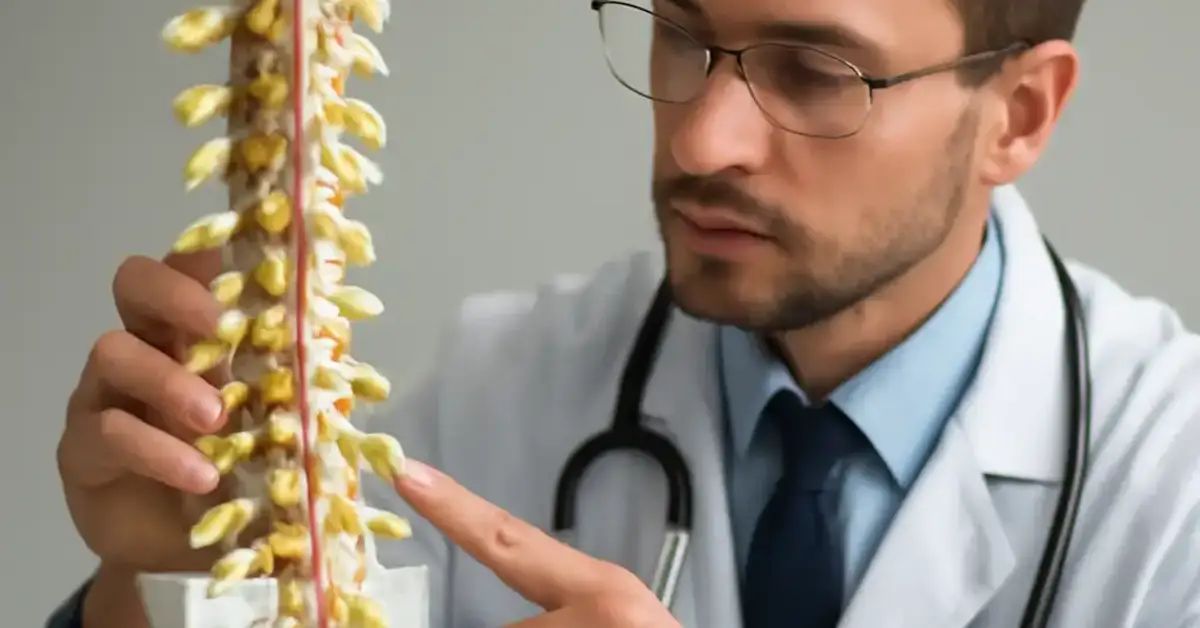
Every medical student, neurology resident, nursing student, and healthcare trainee knows the anxiety of facing a neurology shelf exam or board review. One of the most heavily tested topics is spinal cord syndromes. These syndromes represent the classic injury patterns that arise when specific tracts within the spinal cord are damaged. By learning the anatomy of the corticospinal tract, spinothalamic tract, and dorsal columns, you can predict exactly what deficits a patient will have — and, more importantly, where the lesion is located. This skill is not just academic; it directly impacts clinical decisions about urgent surgical decompression, steroid therapy, and rehabilitation planning. For a related guide, see 8 Simple Ways to Localize Neurological Lesions.
In this article, we break down all 9 neurological syndromes you must know, with clear anatomical correlations, classic presentations, diagnostic pearls, and high-yield exam tips. We also tackle the 20 most common exam questions so you can walk into test day with total confidence.
Complete vs. Incomplete Spinal Cord Injury: The Foundation of All Syndromes
Before diving into individual syndromes, it is crucial to understand the difference between complete and incomplete spinal cord injury. This distinction shapes prognosis and treatment approach.
Complete Spinal Cord Injury
A complete injury means there is no motor, sensory, or autonomic function preserved below the level of the lesion. The American Spinal Injury Association (ASIA) grades this as ASIA A. Recovery of meaningful function is rare, and management focuses on preventing secondary complications such as pressure ulcers, respiratory failure, and autonomic dysreflexia.
Incomplete Spinal Cord Injury
An incomplete injury spares some neurologic function below the lesion level (ASIA B, C, or D). The specific pattern of spared function depends on which tracts remain intact, giving rise to the classic spinal cord syndromes. Incomplete injuries have a better prognosis for functional recovery and often require urgent surgical intervention to preserve remaining function.
The 9 High-Yield Spinal Cord Syndromes You Must Know
Below is a detailed breakdown of each syndrome, including anatomy, presentation, diagnosis, and key exam findings. Bolded LSI keywords highlight essential terms.
1. Anterior Cord Syndrome
Anterior cord syndrome results from damage to the anterior two-thirds of the spinal cord, typically due to ischemia in the anterior spinal artery territory. This artery supplies the corticospinal tract (motor) and spinothalamic tract (pain and temperature). The dorsal columns (vibration and proprioception) are spared because they receive blood from the posterior spinal arteries.
Clinical presentation: The patient presents with bilateral loss of motor function (paralysis or paresis) below the lesion, loss of pain and temperature sensation, but preserved vibration and proprioception. This is a classic neurological syndrome that screams “anterior spinal artery occlusion” until proven otherwise.
Key exam findings: Weakness, spasticity, hyperreflexia, and a positive Babinski sign below the lesion. Pain and temperature sensation are absent, but joint position sense and vibration remain intact.
Common causes: Anterior spinal artery infarction, aortic dissection, surgery involving the aorta, or severe hypotension.
Exam tip: Remember: “Anterior = motor + pain/temp loss, posterior columns spared.”
2. Central Cord Syndrome
Central cord syndrome is the most common incomplete spinal cord injury, typically seen after hyperextension injuries in patients with pre-existing cervical spondylosis. The damage is centered in the central gray matter and the medial portions of the spinothalamic tract and corticospinal tract.
Clinical presentation: The hallmark is disproportionate weakness in the upper extremities compared to the lower extremities. The descending corticospinal fibers to the arms are located more centrally, so they are affected first. Patients may also have a “suspended” or cape-like distribution of pain and temperature loss over the shoulders and upper arms. Sacral sensation is often spared.
Key exam findings: Upper extremity weakness (hands especially weak), lower extremity strength relatively preserved, loss of pain and temperature in a cape distribution, preserved dorsal column function.
Common causes: Hyperextension injury (e.g., fall, motor vehicle accident), spinal stenosis, syringomyelia.
Exam tip: “Central cord = arms weaker than legs.”
3. Brown-Séquard Syndrome (Hemisection)
Brown-Séquard syndrome results from hemisection (one half) of the spinal cord. This classic pattern demonstrates the crossed and uncrossed nature of spinal pathways. The corticospinal tract and dorsal columns are ipsilateral, while the spinothalamic tract crosses at the level of entry.
Clinical presentation: On the same side (ipsilateral) as the lesion: loss of motor function (corticospinal tract) and loss of vibration and proprioception (dorsal columns). On the opposite side (contralateral): loss of pain and temperature sensation (spinothalamic tract) beginning 1–2 segments below the lesion.
Key exam findings: Ipsilateral spastic weakness, ipsilateral loss of vibration and joint position sense, contralateral loss of pain and temperature. This is a high-yield neurological syndrome for exams because the pattern is so specific.
Common causes: Penetrating trauma (knife, gunshot), tumor, demyelinating disease (MS), epidural hematoma.
Exam tip: Draw a cross-section of the spinal cord and label the tracts — it will lock this pattern in your memory.
4. Posterior Cord Syndrome
Posterior cord syndrome is rare and results from damage to the dorsal columns. The corticospinal and spinothalamic tracts are usually spared.
Clinical presentation: Patients have profound loss of vibration and proprioception below the lesion, leading to sensory ataxia (unsteady gait that worsens with eyes closed). Motor function and pain/temperature sensation are preserved.
Key exam findings: Positive Romberg sign, loss of vibration sense, impaired joint position sense, normal strength, and normal pain/temperature sensation.
Common causes: Tabes dorsalis (neurosyphilis), vitamin B12 deficiency (subacute combined degeneration), dorsal column infarct.
Exam tip: Posterior cord = “proprioception problem, not a power problem.”
5. Conus Medullaris Syndrome
Conus medullaris syndrome involves injury to the conus medullaris, the tapered end of the spinal cord at approximately L1–L2. Because the conus houses the sacral segments, this syndrome presents with early and severe autonomic dysfunction.
Clinical presentation: Sudden onset of bilateral saddle anesthesia, loss of bowel and bladder function (urinary retention, constipation), and impotence. Lower extremity motor and sensory loss may be minimal or absent initially. Pain is often less prominent than in cauda equina syndrome.
Key exam findings: Saddle anesthesia, absent bulbocavernosus and anal wink reflexes, loss of voluntary anal sphincter control, preserved lower extremity reflexes early on (later may become hyperreflexic).
Common causes: Tumor, fracture, disk herniation at L1–L2, trauma.
Exam tip: “Conus = cord lesion above the cauda, early bladder/bowel involvement, little leg pain.”
6. Cauda Equina Syndrome
Cauda equina syndrome is a surgical emergency caused by compression of the cauda equina nerve roots below the level of L2. Unlike conus medullaris, this is a lower motor neuron (LMN) injury.
Clinical presentation: Gradual or acute onset of low back pain, bilateral sciatica, saddle anesthesia, lower extremity weakness (often asymmetric), and bladder/bowel dysfunction (urinary retention, overflow incontinence). Pain is often a prominent early feature.
Key exam findings: Flaccid weakness (LMN signs) in the lower extremities, diminished or absent deep tendon reflexes, saddle anesthesia, reduced anal sphincter tone, decreased bulbocavernosus reflex. This is a critical spinal cord injury pattern to recognize immediately.
Common causes: Central disk herniation, tumor, abscess, trauma, spinal stenosis.
Exam tip: “Cauda = nerve roots, LMN signs, severe pain, asymmetric weakness.”
7. Dorsal Column Syndrome (Isolated)
While sometimes grouped with posterior cord syndrome, isolated dorsal column dysfunction can occur as a distinct entity, especially in nutritional deficiencies or early tabes dorsalis. The dorsal columns carry vibration and proprioception.
Clinical presentation: Sensory ataxia, positive Romberg sign, loss of vibration sense in the feet. Strength, pain sensation, and temperature sensation are normal.
Key exam findings: Impaired joint position sense in toes, absent vibration at ankles, Romberg positive, normal strength and reflexes.
Common causes: Vitamin B12 deficiency, syphilis, Friedrich ataxia.
8. Spinothalamic Tract Syndrome (Anterolateral Cord Syndrome)
This is usually seen in combination with other syndromes, but isolated spinothalamic tract damage is occasionally encountered with lateral cord compression (e.g., from a tumor).
Clinical presentation: Contralateral loss of pain and temperature sensation starting 1–2 segments below the lesion. Motor function and dorsal columns are intact.
Key exam findings: Isolated pain/temperature loss on one side, normal strength and vibration sense.
Common causes: Lateral spinal cord tumor, demyelinating plaque.
9. Complete Spinal Cord Transection
Complete transection (ASIA A) is the most severe form of spinal cord injury. All tracts below the level are interrupted, leading to total loss of motor, sensory, and autonomic function.
Clinical presentation: Flaccid paralysis and areflexia immediately (spinal shock), followed over days to weeks by spastic paralysis, hyperreflexia, and autonomic dysreflexia. All sensation is lost below the level, and bowel/bladder function is absent.
Key exam findings: No voluntary motor function, no sensation, absent reflexes initially, then hyperreflexia, Babinski sign present.
Exam tip: “Complete transection = everything gone below the level.”
How Clinicians Localize Spinal Cord Lesions: Neurological Examination and Imaging
Neurological localization is a stepwise process. Start with the pattern of weakness (corticospinal tract), then the pattern of sensory loss (spinothalamic vs. dorsal columns), and finally reflex changes and autonomic signs.
Using the Neurological Exam
A thorough exam includes:
- Motor: Manual muscle testing of upper and lower extremities. Look for upper motor neuron signs (spasticity, hyperreflexia, Babinski) versus lower motor neuron signs (flaccidity, hyporeflexia, atrophy).
- Sensory: Pinprick for pain and temperature (spinothalamic tract), light touch (both tracts), and vibration/proprioception (dorsal columns). A sensory level is a horizontal line below which sensation is altered.
- Reflexes: Deep tendon reflexes, Babinski, bulbocavernosus, anal wink, and cremasteric reflexes help localize the lesion to upper or lower motor neuron.
- Autonomic: Bowel and bladder function, sweating, blood pressure (autonomic dysreflexia suggests high thoracic or cervical lesions).
Imaging Studies
MRI is the imaging modality of choice for spinal cord compression and intrinsic cord lesions. CT myelography is used when MRI is contraindicated. Advanced imaging can identify edema, hemorrhage, tumor, disk herniation, and stenosis. Early imaging is critical in suspected cord compression to prevent irreversible damage.
Comparison Table: Complete vs. Incomplete Spinal Cord Injury Syndromes
| Syndrome | Incomplete? | Motor | Pain/Temp | Vibration/Proprioception | Autonomic |
|---|---|---|---|---|---|
| Anterior Cord | Yes | Lost (bilateral) | Lost (bilateral) | Preserved | Variable |
| Central Cord | Yes | Arms > Legs | Cape loss | Preserved | Variable |
| Brown-Séquard | Yes | Ipsilateral loss | Contralateral loss | Ipsilateral loss | Minimal |
| Posterior Cord | Yes | Preserved | Preserved | Lost (bilateral) | Minimal |
| Conus Medullaris | Yes/No* | Minimal legs | Saddle anesthesia | Variable | Early severe |
| Cauda Equina | Yes | LMN legs (asymmetric) | Saddle anesthesia | Variable | Bowel/bladder |
| Complete Transection | No (ASIA A) | Lost (bilateral) | Lost (bilateral) | Lost (bilateral) | Lost below |
Why Pattern Recognition Matters for Exams and Real Patients
In both medical exam neurology and daily practice, the ability to look at a patient and say, “That’s Brown-Séquard” or “This is central cord syndrome” separates top performers from the rest. Every syndrome has a fingerprint — a specific combination of motor, sensory, and reflex changes. Mastering these patterns helps you answer board questions faster and manage spinal cord injury patients more effectively. For a related guide, see 9 Basic Neuroanatomy Principles You Should Never Forget.
Useful Resources
For deeper study of neuroanatomy exam review and spinal cord pathways, the following resources are highly recommended:
- NCBI Bookshelf: Spinal Cord Syndromes Overview — A comprehensive peer-reviewed resource covering anatomy, classification, and clinical management of spinal cord syndromes.
- Medscape: Spinal Cord Trauma and Injury — Up-to-date clinical guidelines for diagnosis and treatment of spinal cord injuries.
Frequently Asked Questions About Spinal Cord Syndromes
What are the most important spinal cord syndromes for exams?
The most high-yield spinal cord syndromes for medical exams are anterior cord syndrome, central cord syndrome, Brown-Séquard syndrome, posterior cord syndrome, conus medullaris syndrome, cauda equina syndrome, and complete transection. Mastering these will cover the vast majority of board and shelf exam questions.
How do spinal cord syndromes present clinically?
Each syndrome presents with a specific pattern of motor weakness, sensory loss, and reflex changes based on which spinal tracts are damaged. For example, anterior cord syndrome presents with bilateral motor and pain/temperature loss with preserved proprioception, while Brown-Séquard presents with ipsilateral motor and dorsal column loss plus contralateral pain/temperature loss.
What is the difference between complete and incomplete spinal cord injury syndromes?
Complete injury (ASIA A) means no motor or sensory function is preserved below the lesion. Incomplete injuries (ASIA B–D) have some preserved function below the lesion, and the specific pattern determines the syndrome. Incomplete injuries have better prognosis and often require urgent intervention.
How do you recognize anterior cord syndrome ?
Recognize anterior cord syndrome by the triad: bilateral loss of motor function below the lesion, bilateral loss of pain and temperature sensation, and preserved vibration and proprioception. It is most often due to anterior spinal artery infarction.
What causes central cord syndrome ?
Central cord syndrome is most commonly caused by hyperextension injury in patients with cervical spondylosis or spinal stenosis. It can also be caused by syringomyelia. The damage is centered in the central gray matter and medial white matter.
How does Brown-Séquard syndrome present?
Brown-Séquard syndrome presents with ipsilateral loss of motor function and vibration/proprioception (corticospinal and dorsal columns) and contralateral loss of pain and temperature sensation (spinothalamic tract) beginning 1–2 segments below the lesion. It results from hemisection of the cord.
What are the symptoms of posterior cord syndrome?
Posterior cord syndrome causes loss of vibration and proprioception below the lesion, leading to sensory ataxia and a positive Romberg sign. Motor function and pain/temperature sensation are typically preserved.
How is conus medullaris syndrome different from cauda equina syndrome ?
Conus medullaris syndrome involves the spinal cord tip at L1–L2, causing early, severe autonomic dysfunction (bowel/bladder) with minimal leg pain. Cauda equina syndrome involves nerve roots below L2, presents with severe pain, asymmetric LMN weakness, and later autonomic signs. Cauda equina is a surgical emergency.
What are the key exam findings in spinal cord lesions?
Key findings include a sensory level, upper motor neuron signs (spasticity, hyperreflexia, Babinski) for cord lesions, lower motor neuron signs (flaccidity, hyporeflexia) for cauda equina lesions, and specific patterns of weakness and sensory loss that localize the tract involved.
How do spinal cord syndromes affect motor and sensory function?
Motor function is affected via corticospinal tract damage, causing spastic weakness. Pain and temperature sensation are lost with spinothalamic tract damage. Vibration and proprioception are lost with dorsal column damage. The pattern is tract-specific.
What imaging is used to diagnose spinal cord syndromes ?
MRI is the gold standard for imaging the spinal cord and detecting compression, tumors, ischemia, and demyelination. CT myelography is used when MRI is contraindicated (e.g., pacemaker).
What are high-yield spinal cord syndromes for medical exams?
The highest-yield syndromes for medical exams include anterior cord, central cord, Brown-Séquard, posterior cord, conus medullaris, and cauda equina. These appear repeatedly on USMLE, COMLEX, NCLEX, and nursing board exams.
How do you localize spinal cord lesions clinically?
Localize by using the sensory level (determines segmental level), pattern of weakness (UMN vs. LMN), pattern of sensory loss (which tracts are affected), and reflex changes. Cross-sectional anatomy knowledge is essential.
What are emergency signs of spinal cord compression ?
Emergency signs include new-onset bilateral leg weakness, saddle anesthesia, loss of voluntary anal sphincter contraction, urinary retention, and rapid progression of symptoms. These demand immediate imaging and surgical consultation.
How do spinal cord pathways explain clinical symptoms?
The corticospinal tract controls voluntary motor movement; damage causes weakness. The spinothalamic tract carries pain/temperature; damage causes sensory loss on the opposite side. The dorsal columns carry vibration/proprioception; damage causes ipsilateral sensory ataxia. The pattern of deficits directly maps to the tract damaged.
What is the role of the corticospinal tract in spinal cord syndromes ?
Corticospinal tract lesions produce upper motor neuron signs: spastic paralysis, hyperreflexia, clonus, and Babinski sign. The weakness is typically ipsilateral below the lesion (for hemisection) or bilateral (for anterior cord or complete transection).
What is the role of the spinothalamic tract in spinal cord syndromes ?
Spinothalamic tract damage causes contralateral loss of pain and temperature sensation beginning 1–2 segments below the lesion. This is a hallmark of Brown-Séquard syndrome and is also seen in anterior cord syndrome (bilateral loss).
What is the role of the dorsal columns in spinal cord syndromes ?
Dorsal column dysfunction results in ipsilateral loss of vibration and proprioception, leading to sensory ataxia and a positive Romberg sign. It is the defining feature of posterior cord syndrome and is spared in anterior cord syndrome.
What is spinal shock and how does it affect exam findings?
Spinal shock is a temporary loss of reflexes and tone below the level of a complete or severe spinal cord injury. It can last days to weeks. During spinal shock, the patient has flaccid paralysis and areflexia, which later evolves into spasticity and hyperreflexia.
How should I approach studying spinal cord syndromes for exams?
Start with a strong grasp of the cross-sectional anatomy of the spinal cord and the location of the corticospinal, spinothalamic, and dorsal column tracts. Then learn each syndrome’s lesion location and resulting deficits. Use flashcards, draw diagrams, and practice with clinical vignettes. This neuroanatomy exam review approach ensures long-term retention.