Cortical Functions and Associated Deficits Key Takeaways
The human cerebral cortex orchestrates movement, sensation, language, vision, and higher cognition through specialized regions.
- Cortical functions and associated deficits follow a lobe-by-lobe pattern: frontal lobe controls executive function and motor output; parietal lobe integrates sensation and spatial awareness; temporal lobe manages memory and language comprehension; occipital lobe processes vision.
- Primary cortical areas (e.g., motor cortex, sensory cortex) produce focal deficits like paralysis or numbness, while association cortex damage causes complex syndromes such as neglect, aphasia, or agnosia.
- Cortical localization remains a cornerstone of clinical neurology, enabling precise diagnosis of stroke, tumor, trauma, and neurodegenerative disease from bedside examination alone.
Understanding the Cerebral Cortex: Organization and Clinical Relevance
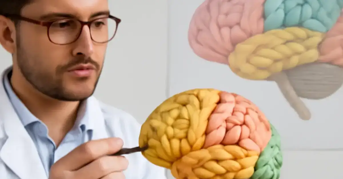
The cerebral cortex anatomy forms the outermost layer of the brain, divided into four lobes: frontal, parietal, temporal, and occipital. Each lobe contains primary areas that execute basic functions and association areas that integrate information for complex behavior. A solid grasp of brain cortex functions allows clinicians to predict deficits from lesion location and plan targeted rehabilitation. For medical students and residents, this knowledge is high-yield for neuroanatomy exam review and daily clinical practice. For a related guide, see 11 Important Brain Structures and Their Clinical Functions.
Frontal Lobe: Executive Hub and Motor Command Center
The frontal lobe houses the primary motor cortex, premotor cortex, prefrontal cortex, and Broca’s area. Its functions span voluntary movement, speech production, executive functions (planning, inhibition, working memory), and personality. Frontal lobe deficits vary by subregion.
Motor Cortex Damage and Contralateral Weakness
The primary motor cortex function is to execute voluntary movement on the opposite side of the body. The precentral gyrus contains a somatotopic map (motor homunculus). Lesions here produce contralateral hemiparesis or hemiplegia, most severe in the face and hand due to their large cortical representation.
Prefrontal Cortex Injury and Executive Dysfunction
Damage to the prefrontal cortex disrupts planning, decision-making, impulse control, and cognitive flexibility. Patients may demonstrate perseveration, poor judgment, apathy, or disinhibition. This syndrome is classic in traumatic brain injury and frontotemporal dementia, often more disabling than motor loss. For a related guide, see 10 Brain Lesions and Their Clinical Presentations Explained.
Broca’s Area and Expressive Aphasia
Broca’s area in the dominant (usually left) inferior frontal gyrus controls speech output. Lesions cause non-fluent aphasia with effortful, telegraphic speech, but preserved comprehension. This is a hallmark of cortical localization in stroke patients.
Parietal Lobe: Sensory Integration and Spatial Awareness
The parietal lobe contains the primary somatosensory cortex in the postcentral gyrus and the superior parietal lobule for higher-order sensory processing. Parietal lobe lesions produce distinct sensory and perceptual syndromes.
Primary Sensory Cortex Damage
Lesions of the sensory cortex damage the postcentral gyrus, causing contralateral loss of touch, pain, temperature, and proprioception. Patients may have difficulty localizing stimuli or recognizing objects by touch (astereognosis).
Neglect Syndrome and Spatial Disorientation
Damage to the right inferior parietal lobule often causes contralateral neglect — the patient ignores the left side of space, draws only the right half of a clock, and may bump into objects on the left. This is a dramatic example of how parietal lobe lesions impair awareness, not just sensation.
Gerstmann Syndrome
Lesions in the dominant angular gyrus produce Gerstmann syndrome: finger agnosia, agraphia, acalculia, and left-right disorientation. This tetrad is a classic cognitive deficits brain injury pattern tested in neuro exams.
Temporal Lobe: Memory, Language, and Auditory Processing
The temporal lobe includes the primary auditory cortex, hippocampus, Wernicke’s area, and extensive association regions. Temporal lobe damage impairs memory, language comprehension, and sound perception.
Hippocampal Lesions and Memory Impairment
The hippocampus is essential for forming new declarative memories. Bilateral damage — as in anoxia or herpes encephalitis — produces anterograde amnesia, the inability to store new facts or events. Unilateral left hippocampal lesions cause verbal memory deficits; right-sided lesions affect visuospatial memory.
Wernicke’s Area and Receptive Aphasia
Wernicke’s area in the dominant superior temporal gyrus enables language comprehension. Lesions produce fluent, jargon-filled speech with poor comprehension. Patients may speak in long runs of nonsensical words, unaware of their errors.
Auditory Processing and Seizures
Primary auditory cortex lesions cause cortical deafness or difficulty localizing sound. Temporal lobe epilepsy often produces auditory hallucinations, déjà vu, and automatisms, reflecting the region’s role in integrating sensory and emotional information.
Occipital Lobe: Visual Cortex and Perception
The occipital lobe contains the primary visual cortex (V1) surrounded by visual association areas. Occipital lobe lesions produce predictable visual field defects and perceptual disturbances.
Primary Visual Cortex Lesions
Damage to V1 in the calcarine sulcus causes contralateral homonymous hemianopia. A complete calcarine lesion yields macular-sparing hemianopia due to dual blood supply. Bilateral occipital damage can cause cortical blindness with preserved pupillary reflexes (Anton syndrome).
Visual Association Area Syndromes
Lesions in extrastriate areas produce higher-order visual deficits: prosopagnosia (difficulty recognizing faces), alexia (inability to read), and visual agnosia (inability to identify objects despite intact vision). These conditions highlight the role of association cortex in interpreting sensory input.
Cortical Association Areas: Integration and Complex Syndromes
Association cortex refers to regions outside primary motor and sensory areas that integrate multimodal information. They are crucial for language, attention, memory, and executive function. Lesions here produce the most diagnostically valuable syndromes in neurology.
Posterior Association Cortex: Parieto-Temporo-Occipital Junction
This region integrates vision, hearing, and touch. Lesions cause syndromes like aphasia, agnosia, and apraxia. For example, a left angular gyrus stroke produces Gerstmann syndrome; right hemisphere damage causes neglect or constructional apraxia.
Prefrontal Association Cortex: Executive Control
The prefrontal association cortex coordinates goal-directed behavior. Cognitive deficits brain injury here manifest as dysexecutive syndrome — impaired planning, set-shifting, and inhibition. Clinicians test this with the Wisconsin Card Sorting Test or verbal fluency tasks.
Primary vs. Association Cortex: Key Differences for Lesion Localization
Understanding the distinction between primary and association areas is fundamental to cortical localization.
| Feature | Primary Cortex | Association Cortex |
|---|---|---|
| Function | Basic processing (e.g., motor execution, sound detection) | Integration and interpretation (e.g., language, spatial reasoning) |
| Examples | Motor cortex (M1), primary somatosensory cortex (S1), primary visual cortex (V1) | Prefrontal cortex, angular gyrus, Wernicke’s area |
| Lesion deficit | Focal, contralateral loss (e.g., weakness, numbness, visual field cut) | Complex syndromes (e.g., aphasia, neglect, agnosia, executive dysfunction) |
| Neuro exam test | Motor strength, sensory mapping, visual field confrontation | Language assessment, clock drawing, verbal fluency, praxis testing |
Clinical Significance: Why Cortical Functions Matter in Practice
For every clinician, from medical students to seasoned neurologists, the ability to localize a lesion based on neurological deficits accelerates diagnosis and management. A patient with right-sided weakness, expressive aphasia, and apathy likely has a left frontal stroke. A patient with left neglect and sensory loss points to a right parietal lesion. These patterns are high-yield for neuroanatomy exam review and daily rounds.
Moreover, understanding brain lobe functions guides rehabilitation goals. A patient with frontal lobe injury benefits from structured routines and executive retraining; someone with temporal lobe amnesia requires environmental cues and repetition. By mastering cortical functions and associated deficits, healthcare professionals improve diagnostic accuracy, patient counseling, and interdisciplinary care.
Useful Resources
To deepen your understanding of cortical functions and associated deficits, consult these authoritative sources:
- Neuroanatomy through Clinical Cases (NCBI Bookshelf) — A comprehensive text linking cortical anatomy to clinical syndromes, ideal for exam preparation.
- American Academy of Neurology (AAN) — Offers guidelines, case studies, and continuous education materials on neurological localization and cortical function testing.
Mastering cortical functions and associated deficits transforms how you approach neurological examination, diagnosis, and patient care. Whether you are preparing for exams or refining your clinical skills, this knowledge remains one of the most practical and rewarding topics in all of neuroscience.
Frequently Asked Questions About Cortical Functions and Associated Deficits
What are the main cortical functions and their associated deficits ?
The main cortical functions include motor control (frontal lobe), somatosensation (parietal), memory and language (temporal), and vision (occipital). Deficits range from contralateral weakness and numbness to aphasia, neglect, amnesia, and visual field cuts, depending on the lobe and specific region affected.
How do different brain lobes affect behavior and cognition?
The frontal lobe governs executive functions, personality, and motor output; the parietal lobe handles spatial awareness and sensory integration; the temporal lobe supports memory, language comprehension, and emotion; the occipital lobe processes visual information. Damage to each produces characteristic changes in behavior and cognition.
What happens when the frontal cortex is damaged?
Frontal cortex damage causes executive dysfunction (poor planning, impulsivity), motor deficits (contralateral weakness), expressive aphasia (if Broca’s area is affected), and personality changes such as apathy or disinhibition, often seen after traumatic brain injury or stroke.
How does parietal lobe injury affect sensation?
Parietal lobe injury impairs contralateral touch, pain, temperature, and proprioception. Patients may lose the ability to recognize objects by touch (astereognosis) or localize stimuli. Right parietal lesions often cause contralateral neglect, where the patient ignores the left side of space.
What are symptoms of temporal lobe dysfunction?
Temporal lobe dysfunction includes memory impairment (especially anterograde amnesia), receptive aphasia (Wernicke’s area), auditory hallucinations, and complex partial seizures. Patients may also exhibit emotional lability or visual agnosia.
How does occipital lobe damage affect vision?
Occipital lobe damage causes contralateral homonymous hemianopia or quadrantanopia. Bilateral occipital lesions lead to cortical blindness with preserved pupillary reflexes. Higher-order deficits like prosopagnosia or alexia occur with extrastriate damage.
What are cortical functions in the brain?
Cortical functions include voluntary movement, somatosensation, vision, hearing, language, memory, attention, executive control, and spatial reasoning. These are distributed across primary and association areas in the frontal, parietal, temporal, and occipital lobes.
How do cortical lesions cause neurological deficits ?
Cortical lesions disrupt the normal processing of sensory, motor, or cognitive information. The deficit pattern depends on the lobe and whether the lesion involves primary cortex (focal loss) or association cortex (complex syndrome). Stroke, tumor, trauma, and infection are common causes.
What are common syndromes of cortical damage?
Common syndromes include contralateral hemiparesis (frontal), neglect and Gerstmann syndrome (parietal), Wernicke’s and Broca’s aphasia (temporal and frontal), homonymous hemianopia (occipital), and dysexecutive syndrome (prefrontal). Each provides clues for lesion localization.
How do you localize cortical brain lesions?
Lesion localization combines history and physical exam. Motor, sensory, visual, and language testing reveals the affected lobe. Neuroimaging (CT/MRI) confirms the location. Knowledge of cortical homunculi and vascular territories aids precise localization.
What cognitive changes occur with cortical injury?
Cognitive changes include executive dysfunction (frontal), memory loss (temporal), language deficits (dominant hemisphere), neglect (right parietal), and visual agnosia (occipito-temporal). The specific cognitive profile guides diagnosis and rehabilitation planning.
How does the cortex control movement and speech?
The primary motor cortex initiates voluntary movement via corticospinal tracts. The premotor and supplementary motor areas coordinate sequences. Broca’s area in the frontal lobe programs speech output. Damage to any of these regions disrupts motor control or speech production.
What are high yield cortical functions for exams?
High-yield functions include motor and sensory homunculus organization, Broca’s and Wernicke’s areas, hippocampal memory circuits, visual pathway, and association cortex syndromes (neglect, Gerstmann, aphasia, agnosia). These are frequently tested in neuroanatomy and board exams.
How do association areas differ from primary cortical areas?
Primary areas perform basic unimodal processing (e.g., detecting light or touch), while association areas integrate multiple inputs for higher functions like language, attention, and planning. Association cortex damage produces complex syndromes, whereas primary cortex lesions cause focal deficits.
What tests assess cortical function clinically?
Bedside tests include motor and sensory exam, visual field confrontation, language assessment (fluency, comprehension, repetition, naming), clock drawing for neglect, frontal lobe tests (verbal fluency, go/no-go), and memory recall. These tests help localize cortical dysfunction.
Can cortical deficits improve over time?
Yes, some recovery occurs through neuroplasticity, especially in younger patients and with intensive rehabilitation. Motor and language functions may partially return, but higher-order deficits like amnesia or neglect often persist chronically. Early intervention improves outcomes.
What is the role of the corpus callosum in cortical function?
The corpus callosum connects the two hemispheres, enabling interhemispheric communication. Lesions cause disconnection syndromes such as alien hand syndrome or alexia without agraphia, revealing how cortical areas depend on cross-hemispheric integration.
How does cortical damage differ from subcortical damage?
Cortical damage produces distinct cortical signs (aphasia, neglect, apraxia, agnosia) that are absent in pure subcortical lesions. Subcortical strokes often cause pure motor or sensory deficits without cognitive changes, though large lesions can affect overlying cortex via edema or disconnection.
What imaging techniques best visualize cortical lesions?
MRI with T1, T2, and FLAIR sequences is the gold standard for detecting cortical lesions, including stroke, tumor, and cortical dysplasia. CT is useful for acute hemorrhage and calcification. Functional MRI (fMRI) maps eloquent cortex preoperatively.
How does epilepsy relate to cortical function deficits?
Epileptic seizures arise from abnormal cortical electrical activity. The seizure semiology reflects the lobe of origin: temporal lobe seizures cause automatisms and déjà vu, frontal lobe seizures produce motor posturing, and occipital seizures cause visual hallucinations. Chronic epilepsy can also impair cortical functions.