Essential Neurology Investigations Key Takeaways
A well-chosen set of essential neurology investigations transforms vague symptoms into a precise neurological diagnosis .
- Master the high yield neurology investigations for exams , including MRI, CT, EEG, and lumbar puncture.
- Understand how brain imaging, neurophysiology, and lab tests complement each other in the clinical neurology workup .
- Learn when to order each test and what the results mean for patient care.
Why Understanding Essential Neurology Investigations Matters for Your Practice
Neurological symptoms can be confusing. Headaches, weakness, numbness, or seizures may stem from dozens of different conditions. Without a systematic approach, clinicians risk missing treatable emergencies like stroke or meningitis. A structured clinical neurology workup built on diagnostic neurology tests helps you narrow the differential diagnosis quickly and safely. For medical students and residents, mastering these tests is not just about passing exams — it is about building the diagnostic reflexes that save lives. For a related guide, see 10 Brain Lesions and Their Clinical Presentations Explained.
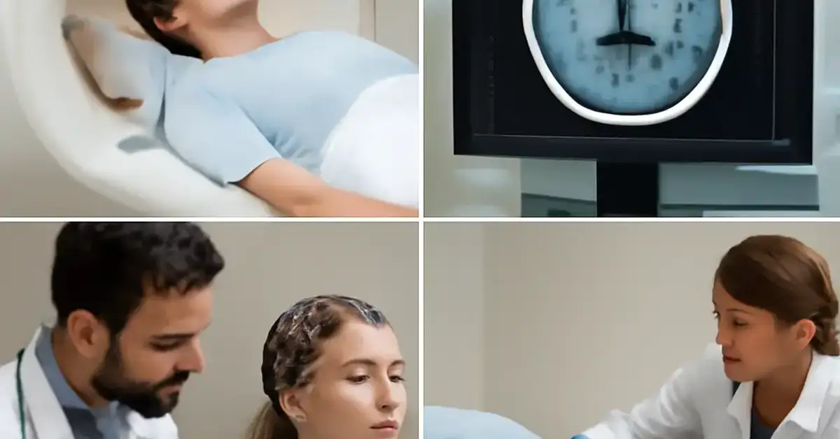
1. MRI Brain Scan – The Gold Standard for Brain Imaging
Magnetic resonance imaging (MRI) uses strong magnetic fields and radio waves to produce detailed images of brain structures. It is the most sensitive test for detecting tumors, multiple sclerosis plaques, encephalitis, and small vessel disease. Brain imaging tests like MRI are often the first step in the neurological diagnosis of unexplained symptoms. When you order an MRI, you can see soft tissue contrast that CT cannot match — making it indispensable for evaluating the brainstem, cerebellum, and white matter.
When Is MRI Used?
MRI is preferred for chronic headache with red flags, suspected demyelinating disease, and evaluation of seizures when structural causes are possible. It also guides surgical planning for brain tumors.
What Do Results Indicate?
Normal MRI rules out many serious conditions. Abnormalities may include mass lesions, enhancement patterns suggestive of inflammation, or white matter hyperintensities typical of small vessel disease. MRI findings often dictate whether you need a lumbar puncture or neurophysiology studies next.
2. CT Scan Brain – Rapid Assessment for Emergencies
Computed tomography (CT) of the brain is the workhorse of emergency stroke investigations. It uses X-rays to create cross-sectional images and is fast, widely available, and excellent at detecting acute hemorrhage, bone fractures, and large masses. In seizure diagnosis tests, CT can identify calcified lesions like neurocysticercosis that may cause epilepsy.
When Is CT Scan Used?
CT is the first-line brain imaging test for acute stroke, head trauma, and suspected subarachnoid hemorrhage. It is also used when MRI is contraindicated (e.g., pacemaker) or unavailable.
What Do Results Indicate?
Hyperdense areas suggest acute blood; hypodense areas may indicate infarction, edema, or chronic ischemia. CT helps determine if a patient is a candidate for thrombolysis in ischemic stroke.
3. EEG Test – Capturing Brain Electrical Activity
An electroencephalogram (EEG) records the brain’s spontaneous electrical activity via scalp electrodes. It is the cornerstone of seizure diagnosis tests and helps classify epilepsy syndromes. EEG can also detect encephalopathy, brain death, and sleep disorders.
How Does EEG Help Detect Seizures?
Interictal epileptiform discharges (spikes or sharp waves) suggest a seizure focus. Ictal recordings capture the actual seizure pattern, guiding medication choice and surgical planning.
What Do Results Indicate?
Normal routine EEG does not rule out epilepsy — sensitivity is about 50%. Prolonged or sleep-deprived studies increase yield. Slowing indicates encephalopathy; specific patterns like burst-suppression are seen in coma or anesthesia.
4. Lumbar Puncture – Accessing Cerebrospinal Fluid
Lumbar puncture (LP) involves inserting a needle into the subarachnoid space to collect cerebrospinal fluid (CSF). It is essential for diagnosing meningitis, encephalitis, subarachnoid hemorrhage (when CT is negative), and demyelinating conditions like multiple sclerosis. Cerebrospinal fluid analysis measures cell count, protein, glucose, and opening pressure, and can detect infections or malignant cells.
What Is a Lumbar Puncture Used For?
LP is done when you suspect CNS infection, inflammatory disease, or leptomeningeal metastases. It is also therapeutic for idiopathic intracranial hypertension (to lower pressure).
What Do Results Indicate?
Elevated white cells with high protein and low glucose suggest bacterial meningitis. Lymphocytic predominance with normal glucose points to viral meningitis. Oligoclonal bands in CSF support multiple sclerosis diagnosis. Traumatic tap causes blood-tinged fluid that clears with tube progression.
5. Nerve Conduction Study – Evaluating Peripheral Nerve Function
Nerve conduction studies (NCS) measure how fast electrical impulses travel along peripheral nerves. They are part of neurophysiology tests used to diagnose carpal tunnel syndrome, peripheral neuropathy, and nerve injuries. The test uses surface electrodes to stimulate the nerve at one point and record the response at another.
How Is Nerve Conduction Study Performed?
A low-intensity electrical pulse is applied to the skin over the nerve. The response is measured in milliseconds and microvolts. It is slightly uncomfortable but does not involve needles.
What Do Results Indicate?
Slow conduction velocity suggests demyelinating neuropathy (e.g., Guillain-Barré). Low amplitude responses point to axonal loss (e.g., diabetic neuropathy). Prolonged distal latency is typical of compression neuropathies like carpal tunnel syndrome.
6. EMG Test – Diagnosing Muscle and Motor Neuron Disorders
Electromyography (EMG) records electrical activity from muscles using a thin needle electrode. It helps distinguish muscle diseases (myopathies) from nerve disorders (neuropathies) and motor neuron diseases like ALS. EMG is often performed together with NCS as part of a comprehensive clinical neurology workup. For a related guide, see 12 Types of Headaches and How to Differentiate Them.
What Is EMG Used to Diagnose?
EMG is used when patients have weakness, atrophy, cramps, or fasciculations. It can detect myasthenia gravis (with repetitive stimulation), myositis, and radiculopathies.
What Do Results Indicate?
Fibrillation potentials and positive sharp waves indicate active denervation. Large, polyphasic motor unit potentials suggest chronic reinnervation. Short-duration, low-amplitude units are typical of myopathy. Repetitive nerve stimulation showing decrement is classic for myasthenia gravis.
7. Blood Tests – The Unsung Heroes of Neurological Diagnosis
While imaging and neurophysiology grab the spotlight, blood tests provide critical information that can confirm or exclude systemic causes of neurologic symptoms. Complete blood count, inflammatory markers (ESR, CRP), autoimmune antibodies (ANA, anti-dsDNA), vitamin B12, thyroid function, and infectious serologies (HIV, syphilis, Lyme) are all important in a comprehensive clinical neurology workup.
What Blood Tests Are Important in Neurology?
For suspected stroke: lipids, coagulation panel, HbA1c. For demyelination: aquaporin-4 antibodies for neuromyelitis optica. For encephalitis: anti-NMDA receptor antibodies. For neuropathy: serum protein electrophoresis for monoclonal gammopathy.
What Do Results Indicate?
Elevated ESR may suggest giant cell arteritis. Vitamin B12 deficiency can cause myelopathy and neuropathy. Positive ANA may point to CNS lupus. Blood tests also guide treatment — for example, ruling out infection before starting immunosuppression.
8. Visual Evoked Potentials (VEP) – Testing Sensory Pathways
VEP is a neurophysiology test that measures the electrical response of the visual cortex to a visual stimulus (usually a checkerboard pattern). It is sensitive for detecting demyelination of the optic nerve, even when the patient has no visual symptoms. VEP is especially useful in evaluating suspected multiple sclerosis.
When Is VEP Used?
Used when optic neuritis is suspected or to find subclinical lesions. It is non-invasive and takes about 30 minutes.
What Do Results Indicate?
A prolonged P100 latency suggests demyelination of the optic nerve. Reduced amplitude may indicate axonal loss. VEP can confirm a clinical suspicion when MRI is equivocal.
9. Cerebral Angiography – Vascular Mapping for Stroke Investigations
Digital subtraction angiography (DSA) is the gold standard for evaluating cerebral blood vessels. It uses contrast dye and X-rays to detect aneurysms, arteriovenous malformations (AVMs), vasculitis, and vascular occlusion. Though MRI and CT angiography (MRA/CTA) are less invasive, DSA provides the highest resolution.
When Is Cerebral Angiography Used?
Used in planning aneurysm coiling or AVM embolization, when there is high suspicion of vasculitis, or when non-invasive tests are inconclusive.
What Do Results Indicate?
Saccular outpouching indicates aneurysm. Early venous filling suggests AVM. Beading of vessels is typical of vasculitis. Complete occlusion confirms ischemic stroke in large vessels.
10. Muscle and Nerve Biopsy – Tissue Diagnosis for Complex Cases
When blood tests, imaging, and neurophysiology are not enough, a small sample of muscle or nerve tissue can be examined under the microscope. This is reserved for suspected vasculitis, mitochondrial disorders, muscular dystrophy, and amyloidosis. It is the definitive way to diagnose certain inflammatory and genetic conditions.
When Is Biopsy Used?
Used when there is progressive weakness of unknown cause, suspected small-fiber neuropathy, or when imaging suggests inflammatory myopathy. Nerve biopsy is typically taken from the sural nerve; muscle biopsy from the vastus lateralis or biceps.
What Do Results Indicate?
Muscle biopsy can show fiber type grouping (denervation), ragged red fibers (mitochondrial disease), or inflammatory infiltrates (polymyositis). Nerve biopsy may reveal demyelination, axonal degeneration, or amyloid deposits.
How Clinicians Choose Diagnostic Neurology Tests
There is no one-size-fits-all approach. The choice of diagnostic neurology tests depends on the clinical presentation, urgency, and suspected localization. A patient with acute hemiparesis gets a CT head stat, then possibly MRI and CTA. A patient with intermittent dizziness may start with MRI brain and inner ear imaging. The high yield neurology investigations for exams are MRI, CT, EEG, LP, NCS, and EMG — these should be memorized cold.
First-Line Tests for Brain Disorders
For suspected structural disease: CT or MRI. For seizure: EEG. For infection: LP. For neuropathy: NCS/EMG. These are the first line tests for brain disorders in most algorithms. Always check with local protocols because some settings use CT head as the first-line for all neurology emergencies.
How Imaging and Lab Tests Complement Each Other
Imaging shows anatomy; lab tests show function. An MRI can show a brainstem lesion, but LP confirms it is inflammatory. EEG can show epileptiform activity, but MRI may reveal the underlying structural cause. Blood work can identify the systemic disease driving the neurological symptoms. Together, they create the complete picture needed for accurate neurological diagnosis and treatment planning.
| Investigation | What It Detects | Best For | Limitation |
|---|---|---|---|
| MRI Brain | Structural lesions, demyelination | Tumor, MS, encephalitis | Slow, contraindicated in some implants |
| CT Brain | Acute hemorrhage, fractures | Stroke, trauma | Less detail in posterior fossa |
| EEG | Seizure activity, encephalopathy | Epilepsy, coma evaluation | Low sensitivity for deep foci |
| Lumbar Puncture | Infection, inflammation, pressure | Meningitis, SAH workup | Invasive, contraindicated with mass effect |
| NCS/EMG | Nerve and muscle function | Neuropathy, myopathy, ALS | Uncomfortable, operator-dependent |
Useful Resources
Deepen your understanding of neurology investigations with these authoritative references:
- NIH: Brain Basics – Imaging and Diagnostic Tests – An excellent government resource explaining how brain imaging and neurophysiology tests work at a foundational level.
- NICE Guideline: Stroke and Transient Ischaemic Attack in Adults – Official UK guidance on when to use CT, MRI, and other tests for stroke diagnosis.
Mastering these ten essential neurology investigations will build your confidence in the clinic and on exams. Whether you are studying for boards or caring for your first patient with a complex neurologic presentation, use this guide as a quick reference. Bookmark it, share it with colleagues, and refer back to it during your next clinical neurology workup.
Frequently Asked Questions About Essential Neurology Investigations
What are the essential neurology investigations ?
The essential neurology investigations include MRI brain scan, CT scan brain, EEG test, lumbar puncture, nerve conduction study, EMG test, blood tests, visual evoked potentials, cerebral angiography, and muscle/nerve biopsy. These tests form the backbone of the clinical neurology workup for most neurologic conditions.
How do doctors diagnose neurological disorders?
Doctors start with a detailed history and neurologic exam, then select from diagnostic neurology tests based on symptoms. Imaging (MRI/CT) localizes structural problems; EEG captures electrical activity; LP analyzes CSF; and NCS/EMG evaluate peripheral nerves and muscles.
What is the role of MRI in neurology?
MRI provides high-resolution images of brain and spinal cord soft tissue. It is essential for detecting tumors, demyelination, stroke, infections, and structural causes of epilepsy. Among brain imaging tests, MRI offers the best detail for most non-hemorrhagic conditions.
When is CT scan used for brain evaluation?
CT is used primarily in emergencies — acute stroke, head trauma, and suspected subarachnoid hemorrhage. It is faster than MRI and excellent at detecting acute blood and bone fractures. It is also used when MRI is unavailable or unsafe.
What tests are used for stroke diagnosis?
Stroke investigations begin with non-contrast CT to rule out hemorrhage, followed by CT angiogram to locate vessel occlusion. MRI with diffusion-weighted imaging can detect early ischemia. Blood tests assess risk factors like cholesterol and clotting function.
How does EEG help detect seizures?
EEG records brain electrical activity. In seizure diagnosis tests, it identifies interictal epileptiform discharges (spikes) that indicate a seizure focus. Ictal EEG captures the seizure pattern, which helps classify epilepsy and guide medication.
What is a lumbar puncture used for?
Lumbar puncture collects cerebrospinal fluid analysis to diagnose meningitis, encephalitis, subarachnoid hemorrhage, multiple sclerosis, and intracranial pressure disorders. It is also used therapeutically to lower pressure in idiopathic intracranial hypertension.
What blood tests are important in neurology?
Key blood tests include CBC, ESR, CRP, vitamin B12, thyroid function, ANA, anti-dsDNA, HIV, syphilis serology, and for specific conditions: aquaporin-4 antibodies (NMO), NMDA receptor antibodies (encephalitis), and serum protein electrophoresis (neuropathy workup).
How is nerve conduction study performed?
A nerve conduction study uses surface electrodes to stimulate a peripheral nerve with a mild electrical pulse. The response is recorded from the muscle or nerve further along the path. It measures conduction velocity and amplitude.
What is EMG used to diagnose?
EMG test is used to diagnose disorders of muscle and motor neurons, including myopathies, neuropathies, radiculopathies, ALS, and myasthenia gravis. It shows denervation, reinnervation, and myopathic changes.
How do clinicians choose neurological investigations?
Clinicians choose based on the localization of symptoms (brain, spinal cord, peripheral nerve, muscle), urgency (acute vs. chronic), and safety (contraindications). High yield neurology investigations for exams are MRI for structure, EEG for function, and LP for fluid analysis.
What are first line tests for brain disorders?
First-line tests include CT brain (for acute emergencies) or MRI brain (for non-urgent structural evaluation). For seizures, EEG is added. For suspected infection, lumbar puncture follows imaging. These are the first line tests for brain disorders in most guidelines.
How do imaging and lab tests complement each other?
Imaging shows anatomy and structural lesions; lab tests (blood, CSF) reveal function, infection, and inflammation. For example, MRI may show a brain lesion, and LP confirms it is demyelinating or infectious. EEG provides functional data when imaging is normal.
What are high yield neurology investigations for exams?
The high yield neurology investigations for exams are MRI brain, CT brain, EEG, lumbar puncture, NCS, and EMG. These appear most frequently in board questions and clinical vignettes. Understand indications, contraindications, and result interpretation for each.
What do neurology test results indicate clinically?
Results from neurology investigations indicate the presence, location, and type of neurologic disease. For example, slow NCS suggests demyelination; fibrillations on EMG indicate denervation; raised CSF protein and low glucose suggest bacterial meningitis; MRI white matter lesions suggest MS or small vessel disease.
Can MRI detect all brain abnormalities?
No. MRI is excellent for structural abnormalities but can miss small lesions, early ischemia (unless diffusion-weighted), and functional disorders like epilepsy without structural cause. That is why EEG and LP are still necessary parts of the clinical neurology workup.
Is CT or MRI better for stroke diagnosis?
CT is best for excluding hemorrhage quickly; MRI with DWI is better for detecting early ischemic stroke. Both are essential stroke investigations. CT is usually done first, then MRI if needed for confirmation or further characterization.
Can EMG be painful?
EMG involves inserting a thin needle into muscles, which can cause mild discomfort similar to a shot. The NCS portion can cause a brief tingling sensation. Most patients tolerate the test well, and the diagnostic information is invaluable for neurophysiology tests.
When is lumbar puncture contraindicated?
LP is contraindicated when there is increased intracranial pressure from a mass lesion (risk of herniation), bleeding disorder, infection at the puncture site, or spinal cord compression. Always review the neurological diagnosis protocol before performing LP.
How long does an EEG take?
A routine EEG takes about 20-40 minutes. Ambulatory EEG can last 24-72 hours. Video-EEG monitoring for presurgical evaluation may require several days. These seizure diagnosis tests provide the data needed to confirm epilepsy and localize the focus.