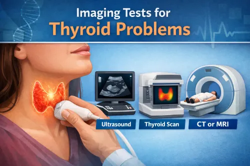
Imaging tests show what the thyroid looks like and how it behaves, complementing blood tests that show how it works. They help doctors assess thyroid size, shape, nodules, and activity so they can distinguish between harmless changes and conditions that need treatment.
You can read your main guide at Imaging Tests and overviews from Cleveland Clinic, Mayo Clinic, and the American Thyroid Association (ATA).
When Are Thyroid Imaging Tests Used?
Doctors usually consider imaging when:
- Blood tests suggest a problem and they need to look at the gland directly
- They feel or see a lump or swelling in the neck
- An incidental thyroid nodule is found on another scan (CT, MRI, carotid ultrasound)
- They are monitoring known nodules, goiter, or thyroid cancer
Imaging helps answer key questions:
- Is the thyroid enlarged (goiter)?
- Are there nodules? If so, how many, how big, and what do they look like?
- Is there anything suspicious for cancer?
- Is the thyroid overactive in certain areas (for example, a “hot” nodule)?
Imaging is usually combined with blood tests described in Blood Tests and the overall pathway in How Thyroid Problems Are Diagnosed.
Thyroid Ultrasound
Thyroid ultrasound is the first‑line imaging test in most people with a thyroid lump or enlarged thyroid.
What It Is
- A non‑invasive, painless test that uses sound waves, not radiation.
- A small probe with gel is moved over the neck while images are taken in real time.
What It Shows
Ultrasound can:
- Measure thyroid size and shape (helping diagnose goiter).
- Detect nodules and describe:
- Size (height, width, depth)
- Composition (solid, cystic, or mixed)
- Edges (smooth or irregular)
- Echogenicity (how bright/dark it looks)
- Microcalcifications and blood flow patterns
These features help classify nodules as low, intermediate, or higher risk, guiding follow‑up and the need for biopsy. Details are described in ATA nodule guidelines and patient pages such as:
- ATA: Thyroid Nodules
- Mayo Clinic: Thyroid nodules – Symptoms & causes
For more on nodules and goiter, see your page Thyroid Nodules & Goiter.
Nuclear Medicine Scans (Radioactive Iodine/Tracer Uptake)
A thyroid uptake scan (sometimes called a radionuclide scan) shows how active the thyroid tissue is.
What It Is
- You swallow a small amount of radioactive iodine (I‑123 or occasionally I‑131) or receive a small injection of a tracer (such as technetium).
- A special camera measures how much tracer the thyroid absorbs and how it is distributed.
What It Shows
The scan can help distinguish causes of hyperthyroidism and characterise nodules:
- Diffuse increased uptake – often seen in Graves’ disease (whole gland overactive).
- Focal “hot” area – a toxic (hot) nodule that is producing excess hormone independently.
- Multiple hot areas – toxic multinodular goiter.
- Low uptake – can suggest thyroiditis, excessive thyroid hormone intake, or certain other causes.
These patterns are described in patient summaries from:
- NIDDK: Hyperthyroidism
- Endocrine Society: Hyperthyroidism
This test is usually ordered when TSH is low and doctors need to clarify the cause of an overactive thyroid. See Hyperthyroidism (Overactive Thyroid) for context.
Fine‑Needle Aspiration (FNA) Under Ultrasound Guidance
Although FNA is technically a biopsy, it is often performed with ultrasound and considered alongside imaging tests.
What It Is
- A very thin needle is inserted into a thyroid nodule to collect cells for analysis.
- Ultrasound guides the needle to the right area, especially for small or deep nodules.
Why It’s Done
- To find out whether a nodule is benign or suspicious/malignant.
- To decide whether surgery or closer follow‑up is needed.
Most nodules are benign, and FNA is the standard way to evaluate suspicious ones, following criteria similar to those in ATA nodule guidelines and described by:
- Mayo Clinic: Thyroid nodules – Biopsy
- Cleveland Clinic: Thyroid Biopsy
Your page Thyroid Nodules & Goiter explains how FNA fits into everyday care.
CT, MRI, and Other Imaging
CT scans and MRI are not routine for simple thyroid problems but are useful in specific situations.
They may be used when:
- A goiter extends behind the breastbone (retrosternal goiter) and doctors need to see how it affects the windpipe or major vessels.
- There is suspected spread of thyroid cancer to lymph nodes or other structures.
- Ultrasound is limited (for example, due to anatomy or very large masses).
PET/CT scans can sometimes detect thyroid incidentalomas (nodules picked up while scanning for other cancers), which then need targeted thyroid evaluation.
These advanced imaging approaches are discussed in oncology‑focused materials such as:
- NCI: Thyroid Cancer—Diagnosis
- StatPearls: Thyroid Cancer
How Imaging and Blood Tests Work Together
Imaging tests do not replace blood tests – they answer different questions:
- Blood tests (TSH, free T4, free T3, antibodies) tell you if the thyroid is underactive, overactive, or under autoimmune attack. See Blood Tests.
- Imaging tests show structure and activity: size, nodules, goiter, or cancer, and whether areas are “hot” or “cold”.
Together, they help your healthcare team:
- Confirm or refine a diagnosis (for example, Graves’ vs toxic nodular disease vs thyroiditis).
- Decide whether a nodule needs biopsy or can simply be monitored.
- Plan treatment – medicine, radioactive iodine, surgery, or observation – as described in Treatment Options for Thyroid Disorders.
Pro Tip for Daily Living
- If you are scheduled for an ultrasound or scan, ask whether you should avoid biotin supplements beforehand, as high‑dose biotin can interfere with some thyroid blood tests (your imaging and blood tests are often coordinated).
- Keep a simple record of imaging dates, results, and key measurements (for example, nodule sizes) so that any future changes can be clearly tracked.
- When you receive a report, ask your provider to summarise it in plain language:
- “Is my thyroid enlarged?”
- “How many nodules do I have, and what is their size/risk level?”
- “Do we need a biopsy or just periodic follow‑up?”
Frequently Asked Questions
Do thyroid ultrasounds or scans expose me to a lot of radiation?
Thyroid ultrasound uses sound waves and involves no radiation at all, while nuclear medicine scans and CT use small, carefully controlled doses that are generally considered safe when medically indicated.
Will I need to stop any medications or supplements before my thyroid imaging test?
Some tests may require you to pause certain medicines or high-dose biotin or iodine supplements for a short time; your healthcare team will give specific instructions in advance.
How long does a thyroid ultrasound or nuclear scan usually take?
A routine thyroid ultrasound often takes around 15–30 minutes, while nuclear medicine scans can take several hours including waiting periods for the tracer to be absorbed.
Is a thyroid ultrasound or FNA biopsy painful?
Ultrasound itself is painless, and FNA biopsy usually causes only mild, brief discomfort at the needle site, similar to a blood test, sometimes with minor soreness afterward.
Can thyroid imaging tests be done during pregnancy or breastfeeding?
Ultrasound is safe in pregnancy and breastfeeding, but nuclear medicine scans are usually avoided or delayed; if urgently needed, special precautions are taken and discussed with you.
How often will I need repeat imaging if I have benign thyroid nodules?
Many benign nodules are monitored with repeat ultrasound every 6–24 months at first, with longer intervals if the size and appearance remain stable over time.
What does it mean if my report says a nodule is “TI-RADS 3” or “intermediate risk”?
These scoring systems combine features like size, composition, and margins to estimate cancer risk and guide whether you need biopsy, closer follow-up, or routine monitoring.
Can thyroid imaging tests tell for sure if a nodule is cancerous?
Imaging can suggest whether a nodule looks more or less suspicious, but a definite diagnosis of cancer usually requires FNA biopsy and, in some cases, surgical pathology.
Will I need someone to drive me home after a thyroid imaging test or FNA?
Most people can go home on their own and resume normal activities after ultrasound, nuclear scans, or FNA, as sedation is rarely used for these procedures.
Can lifestyle factors, like neck posture or weight changes, affect how my thyroid looks on scans?
Body habitus and neck anatomy can influence image quality and how clearly structures are seen, but experienced operators can usually obtain adequate views and interpret results accurately.
Disclaimer: This information is for educational purposes only and does not replace medical advice, diagnosis, or treatment from your own healthcare provider.
Written by: Eden Grace Ramos-Arsenio, RN
Sources: National Institute of Diabetes and Digestive and Kidney Diseases (NIDDK/NIH); Mayo Clinic; NHS; American Thyroid Association (ATA); Cleveland Clinic; Endocrine Society; Thyroid UK; MedlinePlus; peer‑reviewed medical and nursing journals.