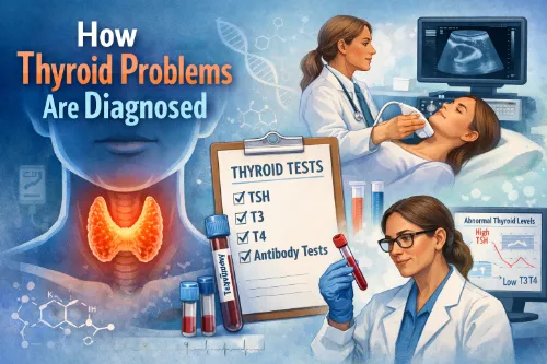
Thyroid problems are usually diagnosed using a combination of your symptoms, a physical examination, and a small number of key tests, especially blood tests and ultrasound. T
his step‑by‑step approach helps your healthcare provider decide whether your thyroid is underactive, overactive, enlarged, or structurally abnormal.
You can read the full overview on your site at How Thyroid Problems Are Diagnosed, along with general patient information from Cleveland Clinic, NIDDK/NIH, Mayo Clinic, and the American Thyroid Association.
How Thyroid Problems Are Diagnosed
Step 1: History and Physical Examination
Diagnosis begins with your story and a focused examination.
Your provider will usually ask about:
- Symptoms such as fatigue, weight changes, temperature intolerance, mood changes, palpitations, bowel changes, and menstrual or sexual health issues.
- Timing and progression – when the symptoms started, whether they are getting worse, and what makes them better or worse.
- Personal and family history of thyroid disease, autoimmune conditions, or thyroid cancer.
- Medications (for example, amiodarone, lithium, immune therapies) and past radiation to the head or neck.
On examination, they may check:
- Your pulse and blood pressure (looking for a slow or fast heart rate).
- The neck, to see if the thyroid is enlarged (goiter) or if there are nodules or tenderness.
- Signs of overactive thyroid (warm, moist skin, tremor, weight loss) or underactive thyroid (dry skin, puffiness, slowed reflexes).
- Eye changes if Graves’ disease is suspected (redness, bulging, or eye movement problems).
This initial assessment guides which tests are needed next.
Step 2: Blood Tests
Blood tests are the cornerstone of thyroid diagnosis. They show how much thyroid hormone is in your blood and how the brain is responding.
Key tests include:
TSH (Thyroid‑Stimulating Hormone)
TSH is made by the pituitary gland and “tells” the thyroid how hard to work.
- High TSH usually means the thyroid is underactive (primary hypothyroidism).
- Low or suppressed TSH usually suggests an overactive thyroid (hyperthyroidism) or too much thyroid medicine.
TSH is often the first test ordered because it is very sensitive to changes in thyroid function.
Free T4 (Free Thyroxine)
Free T4 measures the active form of thyroxine, the main hormone made by the thyroid.
- Low free T4 with high TSH = overt hypothyroidism.
- High free T4 with low TSH = overt hyperthyroidism.
Free T4 helps confirm whether the thyroid is truly under‑ or over‑producing hormone.
Free T3 (Free Triiodothyronine)
Free T3 is sometimes checked when hyperthyroidism is suspected.
- Some people have T3‑predominant hyperthyroidism, where T3 is high but T4 is still in range.
Thyroid Antibodies
These tests help identify autoimmune thyroid disease:
- TPO antibodies (TPOAb) – often raised in Hashimoto’s thyroiditis and sometimes in Graves’ disease.
- Thyroglobulin antibodies (TgAb) – can also be present in autoimmune thyroid disease.
- TSH‑receptor antibodies (TRAb) – often positive in Graves’ disease and help confirm that diagnosis.
Antibodies are particularly useful if the diagnosis is unclear or if there is a strong family or autoimmune background.
Step 3: Imaging Tests
Imaging looks at the structure of the thyroid rather than hormone levels.
Thyroid Ultrasound
Ultrasound is the main imaging test and is:
- Non‑invasive and painless, using sound waves.
- Used to assess size and shape of the thyroid.
- Used to look at nodules – their size, number, appearance (solid or cystic), edges, and blood flow.
Findings help decide:
- Whether a nodule is likely benign or needs closer follow‑up.
- Whether a fine‑needle aspiration (FNA) biopsy is needed to check for cancer cells.
You can read more about structural issues in Thyroid Nodules & Goiter and in patient resources from the ATA and Mayo Clinic.
Radioactive Iodine Uptake Scan (Nuclear Medicine Scan)
This scan shows how active the thyroid tissue is.
- A small amount of radioactive iodine or tracer is swallowed or injected.
- The thyroid is scanned to see how much tracer it takes up and whether the uptake is diffuse (entire gland, often Graves’), focal (a “hot” nodule), or low (thyroiditis or too much thyroid hormone from outside).
Doctors use this test mainly when TSH is low and the cause of hyperthyroidism is unclear.
Step 4: Fine‑Needle Aspiration (FNA) Biopsy
Fine-needle aspiration (FNA) of a thyroid nodule is a quick, minimally invasive test that uses a thin needle to collect cells from the nodule so a pathologist can check for cancer or other abnormalities.
When FNA is recommended
Doctors typically recommend FNA when:
- The nodule is above a certain size (often ≥1 cm if it has suspicious ultrasound features; larger thresholds like 1.5–2.5 cm are used for less suspicious nodules).
- Ultrasound shows concerning features such as microcalcifications, irregular or lobulated margins, marked hypoechogenicity, a taller‑than‑wide shape, or increased internal blood flow.
- The nodule is growing quickly or feels very firm or fixed on exam.
- There is a history of head/neck radiation, strong family history of thyroid cancer, or abnormal lymph nodes in the neck.
Small nodules (<1 cm) may still be biopsied if they look very suspicious or are associated with abnormal lymph nodes, but many low‑risk tiny nodules are just monitored with follow‑up ultrasound.
How the FNA procedure works
- You lie on your back with your neck gently extended, usually with a pillow under your shoulders to better expose the thyroid.
- The skin over your neck is cleaned; a local anesthetic may be injected to numb the area.
- The doctor uses ultrasound to locate the nodule and guide the needle into the precise area.
- A very thin, hollow needle is inserted into the nodule, and the doctor moves it back and forth a few times (often 3–6 passes) to draw up cells and small tissue fragments.
- Each sample is placed on glass slides or in a preservative solution and sent to a pathology lab for microscopic examination.
The whole procedure usually takes about 10–20 minutes and is done as an outpatient; most people resume normal activities the same day.
What the results mean
Pathologists report thyroid FNA results using the Bethesda System, which has six main categories.
- Non‑diagnostic/Unsatisfactory (Category I): Not enough cells or poor sample quality; often leads to a repeat FNA.
- Benign (Category II): No evidence of cancer (for example, colloid nodule or thyroiditis); usual plan is periodic ultrasound follow‑up.
- Atypia of Undetermined Significance / Follicular Lesion of Undetermined Significance – AUS/FLUS (Category III): Some atypical features but not clearly benign or malignant; options include repeat FNA, molecular testing, or surgery depending on risk factors.
- Follicular Neoplasm / Suspicious for Follicular Neoplasm (Category IV): Suggests a follicular‑patterned tumor; many patients are advised to have surgery to remove the lobe for a definitive diagnosis.
- Suspicious for Malignancy (Category V): Strongly suggests cancer but not 100% certain; surgery is usually recommended.
- Malignant (Category VI): Clear features of cancer (most often papillary thyroid carcinoma); treatment typically involves surgery and possibly additional therapies.
Each Bethesda category is linked to an estimated risk of malignancy, ranging roughly from about 0–3% for benign results up to more than 97% for malignant results, which helps guide follow‑up and treatment decisions.
Possible discomfort and risks
- Mild pain, pressure, or bruising in the neck for a day or two is common and usually improves with simple pain relievers.
- Serious complications such as significant bleeding, infection, or damage to nearby structures are rare because the needle is very thin and the procedure is ultrasound‑guided.
Most nodules are benign, but biopsy helps identify the small number that are cancerous or require surgery. You can find more detail in Thyroid Nodules & Goiter and cancer‑focused pages from the American Thyroid Association and National Cancer Institute.
Putting Test Results Together
No single test is interpreted in isolation; your provider looks at the whole picture:
- Overt hypothyroidism: high TSH + low free T4, with symptoms such as fatigue, weight gain, and cold intolerance.
- Subclinical hypothyroidism: slightly high TSH + normal free T4, with mild or no symptoms.
- Overt hyperthyroidism: low TSH + high free T4 and/or T3, with symptoms such as weight loss, tremor, and palpitations.
- Subclinical hyperthyroidism: low TSH + normal T4/T3, sometimes with subtle symptoms or heart/bone risks.
- Euthyroid with nodules/goiter: normal hormone levels but structural changes on exam or ultrasound.
Complex or mixed pictures (for example, after thyroiditis or in pituitary disease) may need repeat testing, additional hormone tests, or referral to an endocrinologist.
When Are Repeat Tests Needed?
Repeat thyroid testing is quite common and plays an essential role in both diagnosis and ongoing care. If your results are borderline or only mildly abnormal—such as a slightly elevated TSH—your doctor may request a follow-up test within a few weeks or months. This helps determine whether levels return to normal on their own or begin to shift further out of range.
For those already receiving treatment, repeat testing helps ensure the medication dose is working as intended. For example, people taking levothyroxine for hypothyroidism or antithyroid drugs for hyperthyroidism need periodic monitoring to keep hormone levels balanced and symptoms under control.
During pregnancy, thyroid monitoring becomes even more important. Hormonal shifts can change your body’s thyroid hormone requirements, so tests are done more frequently to support both maternal and baby health.
Your healthcare provider will recommend the right testing schedule based on your results, treatment plan, and overall health profile—so never hesitate to ask how often you should check your levels and why.
Your doctor will usually explain how often you need testing and what target ranges they are aiming for. More detail on ongoing care is in Long‑Term Monitoring and Treatment Options for Thyroid Disorders.
Pro Tip for Daily Living
Before your appointment, take a few minutes to get organized—it can make a huge difference. Write down your main symptoms and when they began, along with all the medications, supplements, and vitamins you currently take. Don’t forget to note any family history of thyroid or autoimmune conditions; these details help your provider spot potential patterns faster.
If you’ve had thyroid tests done before, bring copies or jot down the dates and results. This allows your provider to look at trends over time, which is much more useful than a single set of numbers.
During your appointment, ask for your specific lab values—TSH, free T4, T3, and thyroid antibodies—and save them in a simple health log, spreadsheet, or phone note.
You don’t need to decode them yourself; the key is having them ready for future checkups or second opinions. Keeping a record empowers you to play an active role in your ongoing thyroid health.
Disclaimer: This information is for educational purposes only and does not replace medical advice, diagnosis, or treatment from your own healthcare provider.
Written by: Eden Grace Ramos-Arsenio, RN
Sources: National Institute of Diabetes and Digestive and Kidney Diseases (NIDDK/NIH); Mayo Clinic; NHS; American Thyroid Association (ATA); Cleveland Clinic; Endocrine Society; Thyroid UK; MedlinePlus; peer‑reviewed medical and nursing journals.